

 PDF
PDF ePub
ePub Citation
 Print
Print


INTRODUCTION
Follicular carcinoma cannot be differentiated from a follicular adenoma based on clinical, cytologic and ultrasonography (US) features. Follicular neoplasms are indeterminate lesions and present a diagnostic challenge to clinicians. Currently, these patients are advised to undergo a hemithyroidectomy and isthmectomy for accurate diagnosis (12). Endoscopic thyroidectomy is considered appropriate for follicular neoplasms because of its outstanding cosmetic results. However, it occasionally leads to unexpected complications such as brachial plexus injury, Horner's syndrome, chyle leaks and operative track seeding (13). Follicular thyroid neoplasm seeding around the operative bed and along the port insertion site is very rare with only 4 cases reported to date. We present a case of follicular thyroid adenoma seeding after endoscopic thyroidectomy for a follicular neoplasm, both in the subcutaneous tunnel of the upper chest wall and in the operative bed.
CASE REPORT
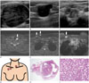
A 14-year-old female visited our hospital because of a palpable neck mass. US using 5–17 MHz linear probe (iU 22; Philips, Bothell, WA, USA) showed a 3.8 cm sized hypoechoic mass with smooth margins in the left lobe of the thyroid gland (Fig. 1A). The diagnosis resulting from fine-needle aspiration biopsy was follicular neoplasm. The patient wanted endoscopic surgery for cosmetic reasons. She elected to undergo an endoscopic gas insufflation left hemithyroidectomy with a bilateral axillo-breast approach (BABA). The final diagnosis of the resected mass was a follicular adenoma. About 3 years after the first surgery, the patient revisited our hospital due to palpable nodules on the right upper chest wall. The nodules were 1–2 cm in size and freely mobile on physical exam. Mammography demonstrated an about 1.7 cm sized, circumscribed, oval, isodensity nodule in the right upper chest wall. On US examination, multiple 0.5–1.9 cm sized circumscribed, oval, hypoechoic nodules were noted in a row along the putative, right, subcutaneous track of the prior endoscopic thyroidectomy, and in the left thyroid bed to level VII (Fig. 1B, C). Magnetic resonance imaging (MRI) (Ingenia 3.0 T; Philips, Best, the Netherlands) performed to determine the exact surgical extent, also showed multiple nodules along the endoscopic track and thyroidectomy bed. These nodules were slightly high to iso-signal intensity on T2 weighted images and iso-signal intensity on T1 weighted images (not shown) with homogenous nodular or peripheral rim enhancement on contrast-enhanced, fat-suppressed axial T1-weighted images (Fig. 1D-F). Schematic illustration showed seeded nodules in the operative bed, infraclavicular area and upper chest wall (Fig. 1G). The patient underwent the surgical resection. The pathological examination of the resected nodules revealed follicular adenoma with the same pattern as the previous hemithyroidectomy specimen (Fig. 1H, I).
DISCUSSION
Recently, endoscopic techniques have been introduced for thyroidectomy instead of classical thyroidectomy (12). Shimazu et al. (4) described the axillo-bilateral-breast approach using both axillary and breast incisions, and Choe et al. (5) included another incision to the contralateral axilla, the BABA. The benefit of endoscopic thyroid surgery is a better cosmetic result compared to conventional open surgery. However, endoscopic thyroid surgery has limits for optimal visualization and complete surgery, and has some complications including rare cases of operative track tumor seeding (5).
Kim et al. (3) reported a patient with papillary thyroid carcinoma recurrence around the operative bed and the subcutaneous tunnel after endoscopic thyroidectomy. Kim et al. (3) suggested that seeding at the trocar site is due to spillage of tumor during tumor manipulation. Traumatic handling of the tumor and inadequate surgical skill are suspected essential factors for port site seeding. Our case is thought to be similar to Kim et al. (3) because tumor seeding occurred along the subcutaneous tunnel of port insertion site. Hur et al. (6) also reported computed tomography findings of follicular thyroid cancer recurrence with multiple, enhancing, bean-sized soft tissue masses around the operative bed and along the port insertion site after endoscopic thyroidectomy. Keiko et al. (7) reported the following mammographic, US and MRI findings of port-site implantation of benign thyroid adenoma after endoscopic thyroidectomy: a polygonal mass with unclear margins on mammography, a heterogenous echoic mass with no interruption of boundary on ultrasound, and a polygonal nodular lesion with clear boundary and peak enhancement in the early phase under contrast on MRI. In our case, two nodules with clear margins were seen on mammography, and US showed well-circumscribed, oval or round hypoechoic lesions. The MRI image showed well-marginated, oval or round nodules with homogenous or rim enhancements along the port insertion site and around the operative bed.
In conclusion, based on US and MRI findings, we report a case of follicular thyroid adenoma, simultaneously seeded along the subcutaneous tunnel of the upper chest wall and around the operative bed. This is a rare, but possible complication after endoscopic thyroidectomy.
 XML Download