

 PDF
PDF ePub
ePub Citation
 Print
Print


INTRODUCTION
Contrast-enhanced head and neck magnetic resonance angiography (MRA) has been widely used for imaging the arterial system of the head and neck. MRA is a noninvasive and safe tool for evaluation of arterial steno-occlusive disease in the head and neck. MRA may eliminate complications associated with conventional catheter-based vascular imaging, such as thromboembolism and iodinated contrast reaction. In addition, MRA is preferred to the time-of-flight (TOF) technique, because it can better depict arterial detail and eliminate the TOF MR angiographic artifacts (12).
An optimally contrast-enhanced MRA of the head and neck is essential for accurate interpretation. In order to acquire optimal images without substantial venous contamination, images should be acquired during the first pass using the peak concentration of the contrast medium (3). The combination of short acquisition times and short contrast infusion times requires precise timing to achieve high-quality angiograms (2). Another important concern is that the angiogram may be degraded by venous stasis or reflux of contrast into the jugular veins, cervical venous plexus, or both. Several previous studies showed higher incidence of venous stasis or reflux with left-arm injections (456). Many factors may contribute to this phenomenon, and anatomical factors in particular may play an important role. In this study we analyzed the causative factors of venous stasis or reflux on contrast-enhanced head and neck MRA.
MATERIALS AND METHODS
Patients
This retrospective study was approved by the Institutional Review Board of our hospital, and the requirement for informed consent was waived. We reviewed patients who underwent contrast-enhanced head and neck MRA from February 2011. Contrast was injected randomly via either the left or the right antecubital vein. The study included patients over the age of 20 years whose medical records documented body weight and height, and whether the patient had hypertension or diabetes mellitus. In addition, patients with a history of cardiac disease, such as heart failure or with abnormality in the mediastinum, such as mediastinal mass or aortic aneurysm were excluded. Patients with poor-quality MRA due to patient movement were also excluded from the study. A total of 150 patients with left-sided injections and 150 patients with right-sided injections were enrolled in this study. The study population consisted of 157 males and 143 females with the mean age of 61.4 years (range, 21–92 years). The body mass index (BMI) was calculated as the quotient between body weight and height squared (kg/m2). Informed consent was obtained before all the studies for the administration of contrast for MRA.
Imaging Technique
Contrast-enhanced head and neck MRA studies were performed using a 1.5-T MR system (Magnatom Avanto, Siemens, Erlangen, Germany). In all cases, examinations were performed using the head and neck coil without breath-hold. A three-dimensional fast low angle shot (3D-FLASH) MRA sequence was used. Image parameters for 3D-FLASH included a repetition time of 2.92 ms, an echo time of 1 ms, a 25° flip angle, a 176 × 384 matrix, a 262.5 × 400-mm field of view, and a 120-mm slab thickness with 104 slices of 1.15 mm effective thickness and no interslice gap. Ten milliliters of gadolinium chelate (Gadobutrol, Gadovist, Bayer Healthcare, Berlin, Germany) were bolus-injected by a MR power injector (Spectris MR injector, Medrad, Warrendale, PA, USA) at a rate of 2 mL/s via an 18-gauge peripheral intravenous catheter. Contrast was injected randomly via either the left or the right antecubital vein with the arm by the patient's side in a neutral position. This was followed immediately by a 20-mL saline flush injected at the same rate. The Combined Application to Reduce Exposure (C.A.R.E., Siemens) bolus technique was used to determine the delay from the start of injection. The acquisition time was 21 s. The source images of all patients were reconstructed in the axial plane to assess the mediastinum.
Image Analysis
MR images were analyzed in consensus by a staff neuroradiologist (E.J.L.) and a third-year radiology resident (D.J.S.). The side of contrast injection, venous stasis, and reflux of contrast into the jugular vein or cervical venous plexus were assessed from MRA studies. We defined venous stasis as the occurrence of a higher signal in the subclavian vein or brachiocephalic vein (BCV) than in the carotid artery. On the axial reconstructions of the source images, we measured the shortest diameter of the right or left BCV and the diameter of the aortic arch. Additionally, we measured the shortest anteroposterior distance between the posterior cortex of the sternum and the anterior cortex of the vertebral body at the level of the shortest diameter of the right or left BCV, and used this measurement for the size of the thoracic cavity. In patients with left or right venous stasis or reflux, we implemented a qualitative image scoring for suboptimal images by using a four-point scale. A score of 0 corresponded to an excellent delineation of the entire arterial system; 1) suboptimal delineation of the ipsilateral subclavian artery according to the venous stasis or reflux; 2) suboptimal delineation of the neck arteries, including the carotid and vertebral arteries, as well as the subclavian artery; 3) suboptimal delineation up to the intracranial arterial system or poor opacification of the entire arterial system. Above the fourth segment, vertebral arteries were defined as intracranial arteries.
Statistical Analysis
All statistical analyses were performed by using the Statistical Package for the Social Sciences software, version 17.0 (SPSS Inc., Chicago, IL, USA). The following variables were evaluated for statistical significance as causative factors for venous stasis or reflux using the χ2 test: differences in the incidence of hypertension, diabetes mellitus, and the side of injection between patients with and without venous stasis or reflux. The mean age, mean BMI, shortest width of the right or left BCV, diameter of the aortic arch, and shortest anteroposterior distance between the sternum and vertebral body between patients with and without venous stasis or reflux were compared using a t-test. The relationship between qualitative image scoring for the suboptimal image and injection side was analyzed using the χ2 test. A p-value less than 0.05 was considered statistically significant. Logistic regression analysis was used to evaluate the relationship between the shortest width of BCV and variables, adjusting for possible confounders, such as patient's age and gender.
RESULTS
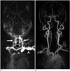
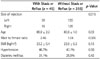
Out of 300 patients, 45 (15%) patients showed venous stasis or reflux (Table 1). Among the 45 patients with venous stasis or reflux, 30 had a left-arm injection, 15 had a right-arm injection (Fig. 1) and the difference was statistically significant (p = 0.015). The mean age of the patients with venous stasis or reflux was 65.9 ± 2.2 years, which was significantly higher than patients without venous stasis or reflux (60.6 ± 1.0 years; p = 0.03) (Table 1). The male-to-female ratio of the patients with venous stasis or reflux was 2.46 (32/13) which was significantly higher than patients without venous stasis or reflux (1.04; p = 0.006) (Table 1). The mean BMI, systemic hypertension, and diabetes mellitus in patients with or without venous stasis or reflux did not differ significantly (Table 1).
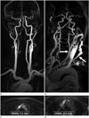
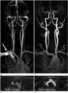
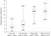
Of 150 patients with left-arm injection, 30 (20%) had venous stasis or reflux (Table 2). The mean age of these 30 patients was 65.1 ± 1.4 years, which was not significantly higher than patients without venous stasis (60.3 ± 2.4 years; p = 0.12) (Table 2). The male-to-female ratio of patients with venous stasis or reflux was 2.33 (21/9), which was significantly higher than that of patients without venous stasis or reflux [0.97 (59/61); p = 0.04] (Table 2). The mean BMI, systemic hypertension, and diabetes mellitus did not differ significantly between the two groups (Table 2). The mean shortest width of the left BCV in patients with venous stasis or reflux (2.96 ± 0.29 mm) was significantly narrower than in patients without venous stasis or reflux (4.80 ± 0.21 mm; p = 0.0001) (Table 2, Figs. 2, 3, 4). After the shortest width of the left BCV was adjusted for gender and age with logistic regression, the width was still significantly associated with venous stasis or reflux (odds ratio = 0.42; 95% confidence interval = 0.27–0.66). There was no significant difference in the mean diameter of the aortic arch (p = 0.21) and the mean shortest anteroposterior distance from the sternum to the vertebral body (p = 0.14) between patients with or without venous stasis or reflux.
In 150 patients with right-arm injection, 15 (10%) patients had venous stasis or reflux. For these 15 patients, there was no significant difference in all variables between patients with or without venous stasis or reflux (Table 3, Figs. 3, 4).
The qualitative image scoring of the left-arm injection group was significantly higher than the right-arm injection group (p = 0.005). The results of the qualitative image scoring for the suboptimal images are summarized in Table 4.
DISCUSSION
Venous stasis or reflux of contrast medium is a major cause of failure in optimally contrast-enhanced head and neck MRA with many related causative factors. Our results showed that 45 of 300 total patients experienced venous stasis or reflux. Among those 45 patients, 30 patients (67%) were injected via the left-arm and 15 patients (33%) were injected via the right-arm. Venous stasis or reflux in the left-arm injected patients was significantly more frequent than in the right-arm injected patients. According to our qualitative suboptimal image score, the left-arm injection group score was significantly higher than the right-arm injection group score, demonstrating that left-arm injected patients with venous reflux had a significantly lower image quality than right-arm injected patients with venous reflux. Similarly, You et al. (7) assessed the retrograde flow of contrast materials from the subclavian vein or BCV, and showed that the left-arm injection had significantly higher scores for retrograde flow of contrast material than did the right-arm injection.
Several previous imaging studies using intravenous contrast, including nuclear medicine studies, CT, and MR imaging also showed that venous stasis or reflux is related to the injection site (567891011). The present results are consistent with previous reports suggesting that the left BCV has a potential for anatomical narrowing. The course of the right BCV is parallel to the ascending aorta. In contrast, the left BCV is longer and less vertical than the right BCV, and must cross between the sternum and the aortic arch or its branches. Therefore, the left BCV can be easily compressed in the retrosternal space, and the compressed left BCV was significantly associated with venous stasis or reflux. In this study, the mean shortest width of the left BCV in patients with venous stasis or reflux was significantly narrower than in patients without venous stasis or reflux, even after adjusting for age and gender.
Furthermore, the left BCV can potentially be compressed in the retrosternal space when the aortic arch shows ectatic change as a consequence of the aging process, systemic hypertension or other factors associated with atherosclerosis. In this study, age was significantly related to venous stasis or reflux, but hypertension, diabetes mellitus and BMI were not. However, other studies reported that hypertension was significantly related to venous stasis or reflux (56). We only checked whether the patient had hypertension by their medical records, whereas these other studies checked patients' current blood pressure. Thus, we suspect that our result with respect to hypertension has some limitations.
According to Tseng et al. (8), decreased retrosternal space was not significantly associated with the dilatation and tortuousness of the aortic arch and its branches; however, Hingwala et al. (5) and Lee et al. (6) reported they were significantly correlated. Therefore, we studied whether the diameter of the aortic arch affected venous stasis or reflux. Among the patients with a left-arm injection, a larger aortic arch diameter was observed in those with venous stasis or reflux, but the difference was not statistically significant. To our knowledge, the diameter of the aortic arch was measured for the first time in our research. Further studies with a larger pool of patients should be conducted to conclude that an accurate relationship between larger aortic arch diameter and venous stasis or reflux. Additionally, we measured the shortest anteroposterior distance between the sternum and vertebral body to determine whether the size of the thoracic cage affected venous stasis or reflux. However, no significant difference was found.
In patients with a right-arm injection, none of the variables showed a significant difference between patients with or without venous stasis or reflux. According to Lee et al. (6), delayed vena cava-pulmonary-aorta transit time, rather than compression of the right BCV, was considered the cause. Absence or incompetence of the internal jugular vein valve has been mentioned in cases with jugular venous reflux (12). The internal jugular valves are the only vascular valves between the right atrium of the heart and the brain (13). According to Anderhuber (14), the internal jugular valves are absent in 13% of the healthy population. Similarly, in a study of 100 autopsy cases (15), 9 cases lacked the venous valve in the left internal jugular vein and 3 cases lacked the valve in the right jugular vein. This factor apparently affects not only the left jugular reflux, but also the right jugular reflux. In the present study, 15 (10%) patients with right-arm injection had venous stasis or reflux. Among these 15 patients, 10 had suboptimal delineation of the ipsilateral subclavian artery contributing to the venous stasis or reflux. In contrast, in the study by Hingwala et al. (5), all patients with venous reflux were injected via the left-arm. However due to the above reasons, venous stasis or reflux and image quality degradation may occur on patient with right-arm injection.
Other factors may also affect venous stasis or reflux. To analyze the causative factors of the venous stasis or reflux in contrast-enhanced head and neck MRA, known anatomical factors affecting the passage of contrast media should be considered. Such factors include the presence of mediastinal masses, aortic aneurysm, vascular anomalies, superior vena cava syndrome, or severe congestive heart failure (12161718). We excluded these factors by evaluating patients' chest radiographs and medical records. In addition, the position of the arm can affect the passage of the contrast media (19). In this study, the patients' arms were placed along the side of their bodies without flexion.
Tanaka et al. (4) found that occlusion of the left BCV disappears at full inspiration, when the retrosternal distance increases. Hernandez et al. (20) demonstrated relaxation of the left brachiocephalic venous compression during inspiration. In this study, MRAs were performed without breath holding. Deep inspiration and breath holding during imaging may also be useful, but these maneuvers may be impractical because of scanning duration.
There are several limitations to this study. First, it was a retrospective design. Second, we did not measure current blood pressure and blood sugar in hypertensive patients and diabetics, respectively. Third, we did not evaluate the insufficiency of the internal jugular valve by using Doppler examination in patients with venous reflux. In further studies, Doppler sonography of the internal jugular vein should be included when assessing the causative factors of the reflux in patients with venous reflux.
In conclusion, compression of the left BCV can lead to venous stasis or reflux that may degrade the quality of contrast-enhanced head and neck MRA. Age is also a major factor contributing to venous stasis or reflux. To acquire an optimal image on contrast-enhanced head and neck MRA, left-arm injection should be avoided, especially in elderly patients.


 XML Download