

 PDF
PDF ePub
ePub Citation
 Print
Print


INTRODUCTION
Acute pulmonary embolism (PE) is found in many clinical settings, and sometimes it can be fatal, with the mortality rate reaching up to 30% if left untreated (1234). Therefore, timely detection of PE is crucial to successful management.
PE is diagnosed clinically based on the modified Well's criteria. The revised version of the American College of Radiology (ACR) appropriateness criteria (2013) recommends that the Well's criteria should be used to calculate the clinical probability of unsuspected PE; however, some of the clinical criteria such as "alternative diagnosis less likely than PE" are considered to be highly subjective and dependent on the physicians' level of experience (5).
At the same time, previously under-recognized cases of unsuspected PE have only recently started to receive much academic attention. In a meta-analysis by Dentali et al., (6) the overall prevalence of unsuspected PE was 1.2% in outpatients and 4.0% in inpatients (6). The negative consequence of unsuspected PE in these patients is as severe as that in symptomatic PE patients (7). Currently, several studies are being conducted to identify the clinical characteristics, outcome, radiologic finding, and proper management of this particular subgroup of patients with PE, which is easily missed in various clinical settings, such as underlying malignancy and hospitalization (5678).
A fatal clinical outcome can be avoided in patients with unsuspected PE when the presence of embolism is incidentally detected through radiologic evaluation while evaluating other lesions using the ACR appropriateness criteria.
When the decision to use the contrast is based on the current ACR appropriateness criteria, a certain proportion of unsuspected PE cases may be further underdiagnosed and "overlooked". The goal of this study is to retrospectively estimate the frequency and clinical characteristics of such overlooked PE, and furthermore, to provide evidence-based recommendations for identifying such overlooked PE cases.
MATERIALS AND METHODS
Patient Selection
The Institutional Review Boards approved this study, and they did not require patient informed consent for retrospective study of case records and CT studies. Through retrospective chart review of patients who were admitted to Seoul St. Mary's Hospital and St. Paul Hospital from January 2007 to July 2014, a total of 939 patients with a final diagnosis of PE were enrolled. These patients included not only the clinically diagnosed PE cases based on the modified Well's criteria and backed up by evidence on chest CT scan, but also unsuspected PE cases that were diagnosed incidentally through a chest CT scan. Patients who did not undergo a chest CT scan, had no emboli on the CT scan, were diagnosed through other imaging modalities such as abdomen CT or cardiac CT, or had a history of chronic PE were excluded. A total of 696 patients were available for further clinical data and image analysis. In patients with multiple episodes of acute PE, the clinical records and images obtained during the initial hospitalization were assessed.
CT Acquisition
CT scanners used during this study period were 16 and 128 multidetector CT systems (Light Speed 16 and Optima 660, GE, Waukesha, WI, USA) at St. Paul Hospital and 16, 64, and 128 multidetector CT systems (Somatom Sensation 16 and 64, Somatom Definition AS, Siemens, Erlangen, Germany) at Seoul St. Mary Hospital. The protocol required contrast material injections at a rate of at least 2–4 mL/sec, at least 100 mL of iodinated contrast material, a section thickness of 5.0 mm or less, 120–140 mA, auto mAs (100–330) depending on the type of scanner, and the use of either bolus timing or bolus tracking software for optimal opacification of pulmonary arteries on pulmonary angiography CT and for ascending aorta on chest CT with contrast.
CT Interpretation
CT scans were retrospectively reviewed by two experienced radiologists working in consensus. The CT prognostic factors–the presence of ventricular septal bowing, the right ventricle (RV)/left ventricle (LV) diameter ratio, and embolic burden score–were evaluated. Ventricular septal bowing was determined if any image showed ventricular septal bowing leftward toward the LV. The diameters of the ventricular cavities were measured on a single axial image obtained at the plane of maximal visualization of the ventricular cavities, and this was usually done at the plane of the mitral valve. The embolic burden was graded by using the previously reported embolic burden scoring system (91011). The score was rated as the number of segmental pulmonary arteries involved, and it was multiplied by two when the thrombus in a vessel was occlusive.
Clinical Data Analysis
Charts were reviewed for demographic data (i.e., age, gender), medical history, and reason for performing chest CT. Laboratory results such as D-dimer level, hemoglobin (Hb) level, and hematocrit (Hct) level, and performance and consciousness status on a scale of 1 to 4 were also obtained. The higher the value, the worse the status of the patient.
In the relevant subgroup of patients, intensive care unit (ICU) admission record, duration of ICU care, death within 3 months of PE diagnosis, and the exact cause of death were recorded.
Simplified ACR Appropriateness Criteria
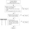
A total of 37 topics, which are the clinical reasons for performing a chest CT scan, were compiled from the ACR appropriateness criteria. The ACR appropriateness criteria include a rating scale from 1 to 9, indicating the level of recommendation for a particular radiological modality from the weakest to strongest. The scale is further divided into 3 sub-groups: the usually not appropriate group (123), the may be appropriate group (456), and the usually appropriate group (789). Specifically with respect to the usage of CT contrast, we systemically re-organized these 37 topics into 4 groups. First, topics with the strongest recommendation were considered. Topics that received at least one 7–9 rating scale for either chest CT with contrast, chest CT with and without contrast, CT angiography, or CT venography chest with contrast were classified into Group A; in contrast, topics with a 7–9 rating scale for chest CT without contrast were classified into Group B. Second, when CT was not recommended at all, if the topics received a 1–3 rating scale for chest CT with contrast, chest CT without contrast, and chest CT with and without contrast, they were classified into Group D. The remaining topics were classified into Group C, uncertainity of contrast usage (Tables 1, 2, Fig. 1).
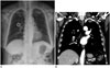
The ACR recommendations for screening of pulmonary metastasis are currently under development. Only in case of primary renal cell carcinoma, the usage of chest CT with contrast is strongly recommended (Group A). In contrast, for primary bone and soft tissue sarcoma, melanoma, testicular cancer, and head and neck cancer, the recommendation advises against the usage of contrast (Group B). For suspected pulmonary metastasis from all other malignancies, patients were classified into Group B (Fig. 2).
Statistical Analysis
We performed statistical analysis using SPSS software (SPPS Inc., Chicago, IL, USA). Continuous variables in each group, such as age and duration of ICU admission, were compared using the one-way ANOVA, and presented as means and standard deviations. Discrete variables, such as gender, ICU admission, death within 3 months, embolic burden score, RV septal bowing, performance score, and consciousness scores were analyzed with the Kruskal-Wallis analysis. Pair-wise comparison was conducted using the Post Hoc tests including Fisher's least significant difference (LSD) and multiple comparison correction and Mann Whitney U test.
RESULTS
Assignment into the Simplified ACR Appropriateness Criteria
Out of the 696 patients, 491 patients were assigned into Group A (70.5%), 104 patients were assigned into Group B (14.9%), 101 patients were assigned in Group C (14.5%), and none of the patients were assigned into Group D. Group A included topics such as evaluation of suspected PE (70%), known primary lung cancer (10%), and deep vein thrombosis (14%). The most frequently found topic in Group B was screening of pulmonary metastasis from primary malignancy such as bone and soft-tissue sarcoma, melanoma, testicular cancer, head and neck cancers, and other cancers (72%). Group B also included topics such as acute respiratory illness in immuno-compromised patients with a positive chest radiograph (15%). Most of the patients in Group C were immuno-competent patients over 40 years of age with acute respiratory illness (90%) (Table 2).
Patient Demographics
Group to group comparison showed that patients in Group C (69.3 ± 14.6 years) were significantly older than those in either Group A (63.3 ± 15.9 years) or Group B (65.8 ± 13.7 years) (p < 0.001). Gender ratio was not statistically different among groups (p = 0.205).
Laboratory Results
The average D-dimer level in Group A patients (8.7 ± 7.7 ug/mL) was higher than that in Group C patients (6.7 ± 6.2 ug/mL) (p < 0.020). The average D-dimer levels in Groups B and C, and Groups A and B were not statistically different. The Hb and Hct levels in Group B patients (11.0 ± 2.0 g/dL, 32.5 ± 5.7%) were statistically lower than those in Group A patients (12.0 ± 2.3 g/dL, 35.6 ± 6.2%) (p < 0.001). The Hb and Hct levels in Group A and Group C (11.8 ± 2.1 g/dL, 35.2 ± 5.9%), and Group B and Group C were not statistically different (Table 3).
Disease Severity and Clinical Outcome
The rate of ICU admission was lowest in Group B (16.3%), compared to either Group A (38.5%) or Group C (36.6%) (p < 0.001). The physical and consciousness status on admission in Groups A and B was not statistically different. However, Group C patients (1.8 ± 0.9) had a higher consciousness status score than Group A patients (1.5 ± 0.8) (p = 0.001); Group C patients (2.0 ± 1.0) also had a higher physical status than Group B patients (1.6 ± 0.9) (p = 0.011) (Table 3). The rate of death within the 3-month follow-up was not significantly different among groups (p = 0.751).
CT Prognostic Factors
The embolic burden score in Group B was the lowest (8.0 ± 5.8), and it was significantly different from that in either Group A (10.7 ± 6.5) or Group C (10.9 ± 6.1) (p < 0.001). Ventricular septal bowing was the least frequently seen in Group B (6.7%), compared to Group A (16.9%) or Group C (21.8%) (p = 0.009). RV dilation was significantly less frequently observed in Group B (8.7%) than in either Group A (22.0%) or Group C (26.7%) (p = 0.003). For all the three factors, the average value in Group A was not significantly different from that in Group C. In composite, evaluation of the three CT prognostic factors showed that Group B had a significantly milder type of PE (Table 4).
DISCUSSION
Retrospective analysis of patients with a final diagnosis of PE showed that a substantial number of our patients may have been overlooked if the ACR appropriateness criteria were applied. Patients with symptomatic PE were more likely to be classified into Group A than into any other group. All patients in Group B and a proportion of patients in Group C are the potential beneficiaries of early radiological diagnosis, which may be more useful when the clinical PE signs are not clearly manifested. In our study, out of the 696 patients, 104 patients (14.9%) were classified into Group B and 101 patients (14.5%) were classified into Group C, respectively.
Especially, a crucial subset of patients in Group B (75 out of 104 patients, 72%) included patients with underlying malignancy, and those who would have not undergone chest CT with the use of ACR appropriateness criteria. They underwent chest CT for screening of pulmonary metastasis, aside from the primary lung and renal cell carcinoma that would have been classified into Group A. The potential relationship between malignancies and venous thromboembolism (VTE), including PE, has been studied quite extensively. Some of the proposed reasons for increased risk of VTE include therapeutic procedure (i.e., surgical procedures, the use of indwelling catheters, local irradiation, and chemotherapy), tumor cell activating factors, and pro-angiogenic cytokines that may directly interact with host vascular and blood cells (12). Such abnormalities were found in as many as 50% of all oncologic patients in that particular study (8).
Further complicating the issue, in oncologic patients, the clinical signs of PE may be less evident and the radiological signs may be different from those in patients with classical PE without malignancy. Bach et al. (12) retrospectively reviewed 3270 patients with PE and reported a rate of 3.9% for unsuspected PE. VTE, PE, as well as unsuspected PE was more commonly detected in oncologic patients (613). Recently, according to Shinagare et al. (14), the rate of symptomatic PE in oncologic patients was 48.9%. Park and Lee (5) also reported a rate of 32.3% for symptomatic PE in the malignancy group and a rate of 64.5% in the control group. This was radiologically explained by the peripheral involvement of PE and lower embolic burden in oncologic patients. According to Park and Lee (5), a retrospective study of 490 patients with PE showed that the subgroup of 155 patients with malignancy presented with preferential involvement of lobar and segmental pulmonary arteries as well as a lower PE index compared to patients without malignancy. This observation is consistent with our finding in Group B patients, mostly cancer patients, who had a lower quantified PE burden and a less chance of ventricular septal bowing and RV dilation.
Oncologic patients have high incidence of unsuspected PE; hence, the clinical significance must not be ignored. Despite the clinically low suspicion for PE, unsuspected PE in oncologic patients was found to be associated with the risk of recurrent VTE, complications and mortality, which were at par with those of symptomatic PE in non-oncologic patients (7). Indeed, as in the case of most of our patients in Group B, these patients with unsuspected PE and underlying malignancies tend to be overlooked under the application of the ACR appropriateness criteria. However, our study showed no significant difference in mortality within the 3-month follow-up among groups; our groups comprised various sets of subgroups and their clinical outcome may have confounded the mortality outcome. This represents a potential area for further revision of the current ACR appropriateness criteria.
In the 2013 revised version of the ACR appropriateness criteria, CT recommendations were summarized for a few common tumors with respect to detecting pulmonary metastases. Patients with bone and soft tissue sarcoma, melanoma, testicular cancer, head and neck cancer were recommended to undergo non-enhanced chest CT, while lung cancer and renal cell carcinoma patients were recommended to undergo enhanced chest CT. The recommendation for usage of CT scan to detect pulmonary metastasis may be difficult to implement in each extra-thoracic malignancy in an everyday clinical setting. However, in a prospective study of 407 oncology patients, the prevalence of unsuspected PE was 6.4% among inpatients, in contrast to a prevalence of 3.4% among outpatients. According to Bach et al. (12), in the study group, PE was found more frequently in patients with primary malignancies of lung, kidney, and breast. Furthermore, previous reports of autopsy series, registry surveys, and epidemiological studies based on hospital discharge data showed that malignancies which are often associated with the development of VTE and symptomatic PE include tumors of the ovary, brain, and pancreas, followed by tumors of the lung, kidney, colon, stomach, prostate, and myeloma (81516171819). The revision of the ACR appropriateness criteria may be accomplished in reference to these preceding study results.
Our study also included Group C, which accounted for 14.5% of patients with PE, who had uncertain clinical evidence for usage of contrast while performing chest CT. The reason for performing chest CT in 84% of Group C patients was acute respiratory illness. Group C patients were older and they displayed worse performance status than Group B patients, and worse consciousness status than Group A patients. Also, evaluation of the rate of ICU admission as well as CT prognostic factors including embolic burden scores, ventricular septal bowing and RV dilatation showed that the patients in Group C had worse clinical presentation and outcome than those in Group A and Group B. As these features of patients with PE were not compared with those in the non-PE control group, it is difficult to state that old age and compromised physical and consciousness status were the risk factors for PE in Group C. Nevertheless, our result suggests that when usage of contrast is uncertain according to the ACR appropriateness criteria and all other factors are equal, patients in old age and with poor performance and consciousness status are more likely to benefit if unsuspected PE was detected radiologically.
There were no patients in Group D. The topics included in Group D were obvious clinical circumstances and a chest plain radiograph was adequate enough to arrive at a clinical diagnosis. Our study revealed that these criteria have been well-understood by physicians and unnecessary chest CT scans were performed.
The present study has the following limitations: first, this study is retrospective in its design, and the CT protocols were slightly varied. There were concerns about variable CT protocols providing different resolutions in evaluation of the embolic burden. However, variable CT protocols are not assumed to affect the evaluation of other prognostic factors, RV dilatation and ventricular septal deviation. Second, the ACR appropriateness criteria itself provides an imprecise rationale for ratings and lack of recommendations for pulmonary screening in most extrathoracic malignancies other than the five tumor classes, mainly sarcoma, melanoma, testicular cancer, head and neck cancer, and renal cell cancer. Clinically important clues such as suspected pleural disease, lymphadenopathy, and vascular invasion are not mentioned. The reason for classifying other malignancies with suspected thoracic metastasis into Group B was based on the established ratings for four extrathoracic malignancies with ACR recommendation to perform non-contrast chest CT scans for evaluating suspected pulmonary metastasis. Third, since the case records were created by various physicians, a certain level of subjectivity was inevitable during the application of our PE-specific simplified version of the ACR criteria. However, the precision of this process was maximized with a careful review of the patients' chief complaint upon admission, progress notes around the date of performing CT, and physicians' notes on the reason for ordering a CT scan. Also, all cases in Groups B and C were not analyzed; therefore, selection bias could not be avoided. Further prospective study of contrast usage in chest CT scans is required based on our study results to evaluate the exact beneficiaries of enhanced chest CT scan. Lastly, the underlying comorbid conditions in individual patients may have confounded the evaluation of clinical severity of PE such as mortality and morbidity. Deaths only due to life-threatening PE were rare, particularly in old patients with significant underlying conditions. It was difficult to delineate the directly PE-induced burden of morbidity and mortality.
Overall, clinical application of our study results requires prudent interpretation. For instance, in cancer subgroups of Group B, the recommendations for non-contrast versus contrast chest scans for the four tumors are 9 vs. 5 for sarcoma, 8 vs. 5 for melanoma, 7 vs. 3 for testicular cancer, 9 vs. 6 for head and neck cancer, where the score range from 4 to 6 indicates "may be appropriate" contrast usage rather than the score range from 7 to 9 which indicates "usually appropriate" contrast usage. While these subgroups of Group B have a stronger recommendation to undergo chest CT without contrast rather than with contrast, such a recommendation is given only in relative terms within one subgroup. In other words, the level of recommendation for contrast usage in some subgroups of Group B does not necessarily indicate low objectivity. Moreover, our study did not analyze more specific clinical outcomes such as mortality and morbidity in these subgroups. However, with the recent increase in reported cases of clinically non-suspected yet fatal PE in oncology patients, a revised and stronger recommendation for contrast usage in certain subgroups is necessary.
Also, in Group C, where the recommendation to perform chest CT scans is uncertain, factors such as old age and physical status may provide useful clinical information for deciding whether or not to use the contrast. Nonetheless, since the usage of contrast has been shown to magnify the amount of DNA damage due to radiation (20), further study should be conducted to clarify a set of specific factors that increase the risk of PE in Group C. Accumulation of such evidence will be helpful in the next revision of the ACR appropriateness criteria.
In conclusion, among cases of PE without clinical suspicion, a substantial number of cases may be overlooked under the application of the ACR criteria related to CT contrast usage. Our retrospective analysis identified a set of patient characteristics and clinical parameters which would help both the clinicians and radiologists in clinical practice.


 XML Download