

 PDF
PDF ePub
ePub Citation
 Print
Print


INTRODUCTION
Neuroblastomas are embryonal tumors that arise from the migrating neuroectodermal cells derived from the neural crest (1). Intracranial neuroblastomas are uncommon and are generally located in the periventricular or supratentorial parenchyma in contrast to olfactory neuroblastomas that originate from the olfactory receptor cells in the nasal cavity (12). To our knowledge, primary sellar neuroblastoma is extremely rare and only 11 cases have been reported since 1979 (13).
We report a case of primary sellar neuroblastoma that showed a solitary sellar mass with supra- and parasellar extension; the tumor mimicked a non-functioning pituitary adenoma or less likely mimicked tuberculum sellae meningioma. We describe the clinical, radiologic, and histopathologic features of our case; additionally, we discuss the current method of treatment for primary sellar neuroblastoma reported in the literature.
CASE REPORT
A 76-year-old man was admitted to our institution for evaluation and treatment of a sellar mass detected after he experienced progressive visual disturbance for 2 months. His medical history was unremarkable except for hypertension. An ophthalmological examination at the time of admission revealed bitemporal hemianopsia and reduced visual acuity that was more severe in the left eye (right 0.5; left 0.25). Endocrine hormone levels were normal, except for slightly increased prolactin (17.7 ng/mL; normal, < 15.2 ng/mL).
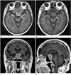
Pituitary MRI at admission revealed a large sellar mass (3.2 × 2.5 × 3.8 cm) with supra- and parasellar extension that caused left posterolateral displacement of the pituitary stalk. The lesion showed homogeneous iso-signal intensity on T1-weighted images, high signal intensity on fluid attenuation inversion recovery images, and relatively homogeneous contrast enhancement. There was no evidence of an abnormality in the floor of the sella turcica or any nasal/ethmoidal bone lesions (Fig. 1).
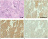
The patient underwent transsphenoidal resection of the tumor. A friable yellow mass was identified upon performing a linear incision over the dura; the mass adhered to adjacent structures. A near total resection was performed, and an intraoperative frozen biopsy indicated a pituitary adenoma. However, histopathologic examination revealed that tumor lobules were separated by dense fibromuscular tissue, and neoplastic cells were small and round with scant cytoplasm, dispersed coarse-to-fine nuclear chromatin, and inconspicuous nuclei. Immunohistochemical results showed strong expression of neural markers including synaptophysin, neural cell-adhesion molecule (NCAM/CD56), and neuron-specific enolase (NSE) (Fig. 2). Immunohistochemical staining was negative for pituitary hormones.
The patient's prolactin levels normalized; however, there was no improvement in his visual disturbance symptoms during the immediate postoperative period. Several imaging examinations were performed to detect any primary or distant lesions; these imaging examinations included neck, abdominopelvic, and chest CT, as well as whole-body positron emission tomography-CT. No evidence of additional pathologic lesions was detected with these modalities.
DISCUSSION
Intracranial neuroblastomas are rare, and they commonly occur in the supratentorial parenchyma or a paraventricular region (4); primary neuroblastomas are extremely rare (5). The origin of this tumor still remains unclear, but Sarwar (5) and Roy et al. (6) suggested that these tumors arise from the ganglion of Locy which grows between the olfactory fossa and the telencephalic vesicle.
Histopathologic examination of sellar neuroblastoma typically reveals homogeneous small cells with round nuclei, obvious fibrillary stroma, and rosette or pseudorosette formations representing a “packed arrangement” of well-differentiated tumor cells. Tumors with a more undifferentiated status show anaplastic hyperchromic small cells with high mitotic activity (7). Immunohistochemical analysis of sellar neuroblastomas typically shows strong expression of neural markers such as synaptophysin, chromogranin, NCAM/CD56, and S-100 protein (7). In the present case, tumor lobules were surrounded by a dense fibrillary stroma, leading to a diagnosis of sellar neuroblastoma. Furthermore, strong expression of synaptophysin, NSE, and NCAM/CD56 was detected on immunohistochemistry.
Dupuy et al. (3) reviewed 9 patients with primary sellar neuroblastomas; their report described that most of these tumors arise primarily in women (77%) with a mean age of 40 years at diagnosis (range, 31 to 57 years). They described that the presenting symptoms were similar to those of a non-functioning pituitary adenoma (98%, bilateral hemianopsia; 66%, hyperprolactinemia; 44%, gonadotropic insufficiency; 11%, hypopituitarism; and 22%, syndrome of inappropriate secretion of anti-diuretic hormone). Our patient was a 76-year-old man; hence, he was the oldest patient among all of the previously reported cases of primary sellar neuroblastomas and one of the infrequent cases of primary sellar neuroblastomas in males.
Radiologic findings of primary sellar neuroblastomas are typically nonspecific on CT and MRI; a sellar mass with or without supra- or parasellar extension can show variable signal intensities on T1- and T2-weighted sequences as well as inconsistent enhancement patterns after gadolinium injection. These findings mimic those of non-secretory pituitary adenoma, tuberculum sellae, or diaphragma sellae meningioma (13). Intraoperative diagnosis can also be equivocal because of the extremely rare occurrence of sellar neuroblastoma and non-specificity of intraoperative findings (1).
Although surgical resection via a transsphenoidal or transcranial approach is the first-line treatment, a uniform postoperative therapy has not yet been established. Dupuy et al. (3) reported the case of a patient who underwent subtotal tumor resection without adjuvant treatment, but was healthy after 3 years. Adjuvant radiotherapy was recommended in the previous studies because of the radiosensitivity of tumors (15). In the present case, a transsphenoidal approach was used for surgical resection; although the tumor was totally excised as per the intraoperative assessment, a follow-up brain MRI performed 2 months later revealed a residual tumor in the posterosuperior aspect of the sella turcica. Therefore, adjuvant radiotherapy was scheduled to prevent tumor progression or metastasis.
In conclusion, primary neuroblastomas located in the sella turcica are extremely rare and they can be difficult to diagnose via radiologic and pathologic examinations. However, primary sellar neuroblastoma should be included in the differential diagnoses of masses located in the sella turcica. Furthermore, understanding primary sellar neuroblastomas in greater detail may assist the radiologists in devising more appropriate multidisciplinary treatment strategies.
 XML Download