

 PDF
PDF ePub
ePub Citation
 Print
Print


INTRODUCTION
Spindle epithelial tumor with thymus-like differentiation (SETTLE) is a very rare malignant thyroid tumor. It arises from ectopic thymus tissue within the thyroid gland or from branchial pouch remnants that differentiate along the thymic line (1). This tumor occurs predominantly in children, adolescents, and young adults (12345). It belongs to a group of cervical lesions that includes ectopic cervical thymoma, ectopic hamartomatous thymoma and carcinoma with thymus-like element (CASTLE) (12345). Although ectopic cervical thymoma and ectopic hamartomatous thymoma are invariably benign, the biologic behavior of SETTLE and CASTLE is generally considered to be malignant. Although SETTLE is originally considered to be a low grade malignant neoplasm because of its indolent growth, it has the potential for late blood-borne metastasis (123456).
To date, several cases of SETTLE have been reported, most of which focused on the histopathologic findings to differentiate SETTLE from other diseases potentially associated with the presence of spindle cells (12345). A few of the reports included imaging studies (26), but to the best of our knowledge, there is no case report in which detailed preoperative imaging features of SETTLE have been described. In addition, there are no case reports of SETTLE in Korean patients. Thus, we report a case of SETTLE with detailed preoperative ultrasonography (US) and computed tomography (CT) features, cytological findings and histopathological results.
CASE REPORT
A 19-year-old woman with a palpable mass on the left side of the neck since the previous 2 months visited a local clinic and was referred to our hospital for evaluation of thyroid nodules. She had no past medical history or risk factors for thyroid malignancy. Laboratory tests revealed normal values on the thyroid function test (T3, 92.64 ng/dL; free T4, 1.25 ng/dL; thyroid-stimulating hormone, 2.23 mIU/L).
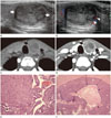
For the evaluation of thyroid nodules, thyroid US was performed by a radiologist with 13 years of experience in using a high-resolution ultrasound instrument (iU 22; Philips Medical System, Bothell, WA, USA) equipped with a 5–12 MHz linear probe. Thyroid US showed a heterogeneous solid nodule in the left lobe (23.6 × 25.1 × 16.7 mm) with benign findings including smooth margin with a hypoechoic halo, oval shape, isoechogenicity, and peripheral vascularity (Fig. 1A, B). No malignant US findings such as marked hypoechogenicity, spiculated margin, microcalcifications, and taller-than-wide shape (7) were observed. In the right lobe, 2 small thyroid nodules with similar findings were identified (less than 5 mm in the largest diameter) (not shown).
For further evaluation, consecutive US-guided fine-needle aspiration (US-FNA) was performed for the largest thyroid nodule in the left lobe. Cytological findings suggested that the nodule was "suspicious for malignancy," and the diagnosis was suspected to be papillary thyroid carcinoma (PTC), because some tight clusters of cells showing papillary structures with nuclear clearing and occasionally pseudo-inclusion like features were observed.
For the purpose of preoperative nodal staging, sequential neck CT was performed using a 64-channel multi-detector CT scanner (Aquilion One; Toshiba Medical Systems, Otawara, Japan) with injection of contrast medium [130 mL iopamidol (Pamiray 370); Dongkook Pharmaceutical, Seoul, Korea; 2.5 mL/s; delay 40 seconds]. On neck CT, 3 solid thyroid nodules corresponding to those on thyroid US were identified. The left thyroid nodule showed heterogeneous low attenuation [mean attenuation value; peripheral portion of the tumor 36 ± 9.91 Hounsfield units (HU), central portion of the tumor 9 ± 6.73 HU, ipsilateral parenchyma 183 ± 6.38 HU] and no calcification on the non-enhanced axial CT image (Fig. 1C), and smooth margin and heterogeneous enhancement (mean attenuation value: peripheral portion of the tumor 118 ± 21.18 HU, central portion of the tumor 24 ± 8.71 HU, ipsilateral parenchyma 212 ± 39.94 HU) on the 40-second delayed contrast-enhanced axial CT image (Fig. 1D). There were no other malignant findings such as adjacent soft tissue invasion or suspicious lymphadenopathy.
On the basis of the malignant cytology result, total thyroidectomy and nodal dissection of the left central nodes were performed. A histopathological section of the left thyroid nodule showed a biphasic pattern with predominant spindle cells arranged in variable papillae, tubules, and glomeruloid structures and a minor glandular mucous component lined by respiratory-type epithelium (Fig. 1E). The mass was encapsulated by a thick fibrous capsule. There was a pseudocyst composed of a large cystic space and fibrinous material deposition in the central portion of the left thyroid nodule (Fig. 1F). The final diagnosis was SETTLE in the left lobe and hyperplasia of the 2 nodules was observed in the right lobe. The patient is free of the disease during a follow-up period of at least one year after surgery.
DISCUSSION
Initially, SETTLE tumor was named spindle cell tumor with mucous cysts, ectopic cervical thymoma or malignant teratoma of the thyroid gland (12). Chan and Rosai (1) first reported the unifying concept of SETTLE tumor of the thyroid in 1991. They postulated that SETTLE tumor arises from ectopic thymus tissue in the thyroid gland or from branchial pouch remnants. Although SETTLE grows slowly, metastasis occurred in 35% of the reported cases of SETTLE during the course of the disease (3). Interestingly, with more than 5 years of follow-up after diagnosis, 71% of patients with SETTLE showed metastasis (4). The common sites of metastasis are lungs, kidney, mediastinum, lymph nodes, liver, and vertebrae (1234). The malignant potential of this tumor might be underestimated because of the lack of long-term follow-up. Thus, long-term follow-up (> 10 years) is needed even in patients with an apparent cure. No guideline regarding the optimum treatment for this tumor has been established owing to the paucity of data. Surgical resection is the mainstay of therapy for SETTLE. There is no known role for adjuvant chemotherapy or radiotherapy.
According to the United States National Center for Biotechnology Research (PUBMED database), only 42 cases of SETTLE have been reported in the form of case reports or case series. Histologically, many investigators have reported the biphasic pattern of SETTLE with predominant spindle cells arranged in variable papillae, tubules, and glomeruloid structures and a minor glandular mucous component (12345). However, little is known about the imaging features of SETTLE; only 2 CT images were presented in previous reports (26). Hence, information pertaining to the imaging characteristics of these tumors, such as the "solid" and "heterogeneous" features, is limited (26).
In the present report, SETTLE showed benign findings on US. The reason for this occurrence is not clear. The tumor was encapsulated by a thick fibrous capsule on histopathological examination, and this might have corresponded to the smooth margin with a hypoechoic halo on US. On macroscopic examination of SETTLE, our gross findings with "a smooth and well-defined edge surrounded by a capsule" were similar to those in other case reports (268), except for a case with an infiltrative border at a very advanced stage in a 12-year-old girl (5). In our case, the tumor showed heterogeneous enhancement on CT and a heterogeneously enhancing peripheral portion and a less enhancing central portion were detected. Although pathologically, direct matching of each portion of the tumor with different degrees of enhancement is limited, we suggest that the biphasic pattern of SETTLE with predominant spindle cells arranged in variable papillae, tubules, and glomeruloid structures and a minor glandular mucous component may have a role in heterogeneous enhancement of the tumor. According to the review of microscopic specimen, the less enhancing central portion of the tumor was a pseudocyst with fibrinous material deposition, and it was thought to be caused by an old hemorrhagic change of unknown origin. Unlike thyroid US, CT features of malignant and benign thyroid nodules have not been established. Recently, Kim et al. (9) suggested that there was a significant difference between malignant and benign solid thyroid nodules in the degree and pattern of nodular enhancement, but other CT features showed no significant differences. The prevalence of homogeneously decreased enhancement in malignant nodules (93.0%, 80/86) was higher than that in benign nodules (7.0%, 6/86) (9). However, further studies assessing the CT features of thyroid nodules are required.
On cytological examination, our case mimicked PTC. Because the findings of US-FNA reflect only those of the targeted point, SETTLE can be confused with other tumors that show the characteristics of the targeted point due the biphasic histological pattern of SETTLE (12345). In addition, the fact that this tumor can show a papillary architecture and the presence of pseudo-inclusions in the preparation can mislead the clinicians to the diagnosis of PTC (8), similar to our case. Differential diagnoses of SETTLE based on these discordant imaging-cytology findings include hyalinizing trabecular tumor (HTT), which is an essentially benign tumor. Despite benign findings on US, HTT can be misinterpreted as PTC on cytological examination because of intranuclear grooves and intranuclear inclusions (10). SETTLE may also be confused with the follicular variant of PTC, which shows less malignant findings on US than does classic PTC.
In summary, we reported a case of SETTLE, which showed benign findings on US, heterogeneous enhancement on CT, and mimicked PTC on cytological examination. We suggest that SETTLE should be included in the differential diagnoses when heterogeneous solid thyroid nodules in children, adolescents, and young adults show benign findings on US and mimic PTC on cytological examination.
 XML Download