

 PDF
PDF ePub
ePub Citation
 Print
Print


Abstract
Periureteral varices are rare, and periureteral varices with accompanying pyelitis have been even more rarely reported. Our patient with flank pain was diagnosed with pyelitis because her CT scan showed thickening and enhancement of the ipsilateral renal pelvic wall and her clinical manifestations and laboratory results were well correlated with the diagnosis. Moreover, the author diagnosed periureteral varices because periureteral, tortuous, enhancing vascular structures were detected and 3-dimensional rendering technique of the CT scan showed that periureteral varices were connected to the ipsilateral renal vein. We experienced a case of periureteral varices with accompanying pyelitis, which was definitely confirmed by using the 3-dimensional rendering technique of the CT scan. Therefore, we report the case along with a brief review of the literatures regarding periureteral varices.
Figures and Tables
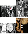
Fig. 1
Periureteral varices in a 70-year-old woman.
A, B. Corticomedullary phase sagittal CT scan (A) and multiplanar curved reformatted image (B) reveal tortuous, tubular venous structures (arrows) along the left ureter. Mild thickening with enhancement of the left renal pelvic wall suggesting pyelitis is noted (red arrows).
C. Maximum intensity projection image shows periureteral varices connected to the Lt. renal vein (arrows).
D. Volume rendering image demonstrates periureteral varices (arrows).
References
1. Heal MR. Ureteral varicosities--a cause of the corkscrew ureter. Br J Surg. 1970; 57:274–276.
2. Weiner SN, Bernstein RG, Morehouse H, Golden RA. Hematuria secondary to left peripelvic and gonadal vein varices. Urology. 1983; 22:81–84.
3. Wendel RG, Crawford ED, Hehman KN. The "nutcracker" phenomenon: an unusual cause for renal varicosities with hematuria. J Urol. 1980; 123:761–763.
4. Kim HS, Park JW, Won IS, Shin KC, Kim S, Yang J, et al. A case of periureteral varices with nutcracker syndrome diagnosed by intravenous pyelography. Korean J Nephrol. 2009; 28:142–145.
5. Trambert JJ, Rabin AM, Weiss KL, Tein AB. Pericaliceal varices due to the nutcracker phenomenon. AJR Am J Roentgenol. 1990; 154:305–306.
6. Ali Khan S, Jayachandran S, Desai PG, Bonheim P. Renal colic, a presenting symptom of pelviureteric varices. Int Urol Nephrol. 1985; 17:11–14.
7. Martelli A, Vitullo F. Microscopic hematuria due to peri-ureteral varices. Urol Int. 1970; 25:457–465.
8. Johnson PT, Halpern EJ, Kuszyk BS, Heath DG, Wechsler RJ, Nazarian LN, et al. Renal artery stenosis: CT angiography--comparison of real-time volume-rendering and maximum intensity projection algorithms. Radiology. 1999; 211:337–343.
9. Cuéllar i Calàbria H, Quiroga Gómez S, Sebastià Cerqueda C, Boyé de la Presa R, Miranda A, Alvarez-Castells A. Nutcracker or left renal vein compression phenomenon: multidetector computed tomography findings and clinical significance. Eur Radiol. 2005; 15:1745–1751.
 XML Download