

 PDF
PDF ePub
ePub Citation
 Print
Print


Tenosynovial giant cell tumor (TSGCT) is classified as a benign fibrohistiocytic tumor according to the World Health Organization (WHO) system of classification of bone and soft-tissue tumors. The proposed causes and pathogenesis include inflammation, trauma, toxin, allergy, and clonal chromosomal abnormalities (1). Jaffe et al. (2) described this condition as pigmented villonodular proliferative lesions arising from the synovium, bursa, or joint. Based on the growth characteristics of TSGCT, it can be divided into two forms: localized type and diffuse type. Localized-type TSGCT encompasses giant cell tumors of the tendon sheath and localized pigmented villonodular synovitis (PVNS), whereas diffuse types include conventional PVNS and diffuse giant cell tumors (3). TSGCT rarely involves the bursa, and only a few exceptional cases of TSGCT arising from the pes anserine bursa have been reported (4). In TSGCT, bone erosion is not an uncommon associated finding, especially in tumors of the tendon sheath of the finger or toe or in the joint space. However, only one previous case of associated bone erosion in TSGCT of the pes anserine bursa has been reported (5). Herein, we report an exceptional case of TSGCT of the pes anserine bursa with bone marrow extension on magnetic resonance imaging (MRI). This case report was approved by the Institutional Review Board of our hospital, and the requirement for obtaining informed consent was waived.
CASE REPORT
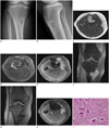
A 41-year-old female patient presented with a slow-growing mass on the medial aspect of her right proximal leg for the past three months. She did not complain of pain, functional disability, loss of motion, or locking of the joints. She denied any history of trauma or radiotherapy. Physical examination revealed a firm, palpable, non-tender 4.0 × 4 .0-cm mass on the medial aspect of the right proximal knee at the level of the insertion site of the pes anserinus and it appeared to be fixed to the tibia. The right medial knee did not exhibit warmth, erythema, or induration. Range of motion of the knee was not limited. Radiographs demonstrated a well-defined, juxtacortical, lobulated soft tissue mass with an eccentric geographic osteolytic lesion with a thick sclerotic rim in the proximal metaphysis of the tibia. The bone marrow adjacent to the mass was intact (Fig. 1A, B). Tumor matrix calcification was not observed. Differential diagnosis included juxtacortical benign soft tissue masses, such as periosteal ganglion cyst, hemangioma, and periosteal chondroma. The MRI was suggestive of a 4.9 × 3.0-cm-sized lobulated solid mass at the insertion site of the pes anserinus conjoined tendon. This tumor exhibited an iso- to hypointense signal on T1-weighted images (Fig. 1C) and a hypo- to hyperintense signal on T2-weighted images (Fig. 1D) as compared to the surrounding muscle and it showed heterogeneous enhancement (Fig. 1E, F) and scattered low signal dots, mostly located peripherally, on all pulse sequences (Fig. 1C-E). Gradient echo coronal imaging demonstrated peripherally located, low signal intensity dots in the mass (Fig. 1G). The tumor had invaded the bone marrow of the tibia, but there was no abnormal signal intensity in the surrounding bone marrow (Fig. 1C), and there was no knee joint involvement. The tendons of the semitendinosus, gracilis, and sartorius muscles were encased by the tumor (Fig. 1H). The preoperative diagnosis was a benign soft tissue mass with internal hemosiderin located in the pes anserine bursa, considered as a tenosynovial giant cell tumor, and chronic bursitis with internal hemorrhage.
Total mass excision was performed. Histopathological examination of the specimen showed a benign lesion with osteoclast-like multinucleated giant cells and sheets of mononuclear cells accompanied by hemosiderin deposits (Fig. 1I). There was no atypia, mitosis, or necrosis. These findings were compatible with a clinical diagnosis of TSGCT of the pes anserine bursa. After surgery, the recovery was propitious. There has been no tumor recurrence and the patient was disease-free without any functional limitations at the 1-year follow-up visit.
DISCUSSION
Since the nomenclature of PVNS was first proposed by Jaffe et al. (2) in 1941, several terms with a similar meaning such as diffuse-type giant cell tumor of the tendon sheath arising from the bursa, pigmented villonodular bursitis (PVNB) and pigmented villonodular tenosynovitis were used before the revision of WHO Classification of Tumors in 2013. For example, a previous report used the term PVNB mixed with diffuse giant cell tumor of the pes anserine bursa (6). Therefore, in order to avoid confusion over the terms, we used the term diffuse-type TSGCT in this report according to the revised WHO classification.
TSGCTs originate from the synovial tissue in various regions of the body, and they can be classified into localized type and diffuse type according to the shape and extent, regardless of the intra- or extra-articular location (37). The localized type is a well-circumscribed mass, whereas the diffuse type is an ill-defined, infiltrative mass or it involves the whole joint space. Localized lesions are more indolent in general (3), while diffuse-type TSGCTs are defined by invasive, extra-articular disease, regardless of whether they arose from the soft tissue or the joint. Unlike localized TSGCTs, diffuse-type TSGCTs usually infiltrate the adjacent soft tissue and erode the bone. Most cases of extra-articular diffuse-type TSGCTs represent extra-articular extensions of primary intra-articular disease (3); however, because of their infiltrative growth pattern, it is often difficult to determine the origin. Due to its invasiveness, diffuse-type TSGCT has a 33 to 50% risk of recurrence and it frequently exhibits multiple recurrences. A histologically benign diffuse-type TSGCT can metastasize with multiple recurrences, but this occurs rarely (3). In our case, the tumor was located in the extra-articular pes anserine bursa and had an infiltrative margin; hence, it was classified as extra-articular diffuse-type TSGCT.
Extra-articular diffuse-type TSGCT is extremely rare and tends to occur in the same locations as intra-articular conventional PVNS. Involvement of a true bursa without articular communication is the least common in extra-articular diffuse-type TSGCT. A small number of such cases involving the bursa around the knee, such as suprapatellar and popliteal bursa, have been reported, but a review of the literature revealed only a few articles about extra-articular diffuse-type TSGCT of the pes anserine bursa. Patient age in TSGCT of the pes anserine bursa ranges from 11 to 63 years, with a mean of 31 years and without any observed patterns in terms of sex or side. Only one patient presented with a history of trauma (4).
The most common radiographic appearances of TSGCT are soft tissue swelling, joint effusion and bone erosion without joint space invasion (7). However, diagnosis of TSGCT based on radiography is difficult because the findings are non-specific. Furthermore, bony erosion is uncommon in TSGCTs of the bursa. Sonographic findings of TSGCT are non-specific, and they have been described as both hypoechoic and hyperechoic in previous reports. However, most TSGCTs show substantial blood flow on color Doppler sonography due to hypervascularity (78). MRI typically reveals a well-defined soft-tissue mass with low to intermediate signal intensity compared to muscle tissue on T1-weighted images and predominantly low signal intensity on T2-weighted images. Areas of decreased signal intensity on all pulse sequences are characteristic of TSGCT due to the presence of hemosiderin and are manifested more in the periphery of the lesions. These areas are associated with the magnetic susceptibility effect and they are more pronounced on gradient echo images. After contrast administration, strong homogeneous enhancement is usually seen (17).
Although a review of TSGCT of the pes anserine bursa reported one among 12 cases with mechanical pressure on the tibia, a direct connection between extra-articular diffuse-type TSGCT and bony change was not proven because only radiographic and CT findings were reported (45).
In our case, radiography showed an eccentric geographic osteolytic lesion with a thick sclerotic rim, and a juxtacortical, lobulated soft tissue mass. The size of the soft tissue mass was larger than the bony involvement, suggesting a benign juxtacortical soft tissue mass. MRI showed a large lobulated soft tissue mass with an infiltrative margin located in the pes anserine bursa. This tumor directly invaded the bone marrow of the tibia through cortical destruction. The lesion exhibited low signal intensity dots related to hemosiderin on all pulse sequences and blooming artifact on gradient echo images. The infiltrative contours and bone involvement seen in our case are common findings of hemangioma. However, the signal of hemangioma on T2-weighted imaging and the enhancement on contrast-enhanced imaging are brighter than those seen on MRI in our case. Also, hemangioma exhibits internal low signal intensity on T2-weighted imaging that appears to be septal and nodular in shape, rather than in the form of scattered dots, as seen in our case (9). Considering the typical location of the pes anserine bursa and the characteristic signal of hemosiderin, TSGCT and chronic bursitis with internal hemorrhage were included in the differential diagnosis. Chronic bursitis with internal hemorrhage appears as a cystic mass with internal hematoma with various phases or fluid-fluid levels, and usually exhibits a hemosiderin rim, and not scattered hemosiderin dots (10). Also, chronic bursitis usually appears as a well-defined mass, which differs from the irregular contours seen in our case.
Differentiation between benignity and malignancy is based on cellular pleomorphism and mitotic activity on histologic examination (47); however, many malignant TSGCTs are radiation-associated tumors, which occur many years after radiotherapy for uncontrolled PVNS (3). Our case was confirmed to be that of a benign diffuse-type TSGCT. Treatment consists of careful and complete local excision and post-operative radiotherapy. Adequate initial local excision may effectively limit the risk of local recurrence.
In conclusion, extra-articular diffuse-type TSGCT of the pes anserine bursa is extremely rare, and it has only been reported once before to occur along with bone erosion on radiography and CT without MRI. No previous reports have shown direct bone involvement with bone marrow extension of extra-articular diffuse-type TSGCT of the pes anserine bursa. While TSGCT can present in very unusual locations like the pes anserine bursa and can have unusual imaging findings like bone marrow extension, characteristic MRI findings, including the presence of hemosiderin, can help to diagnose extra-articular diffuse-type TSGCT.
 XML Download