

 PDF
PDF ePub
ePub Citation
 Print
Print


Abstract
Purpose
To investigate radiologic findings of intraosseous lipoma on plain radiograph and magnetic resonance imaging (MRI).
Materials and Methods
Twenty-seven radiologically or pathologically confirmed intraosseous lipomas of long bones were included in the study. The size, involved bone and site, bone destruction pattern, border, internal calcification, bony expansion, cortical disruption and endosteal erosion were retrospectively analyzed on plain radiograph. The cases were classified into three stages based on calcification and cystic change. Eccentricity, margin, signal intensity of internal fatty portion, and presence of enhancement were analyzed.
Results
Twelve tumors were located in femur and 8 in humerus. Proximal metaphysis was the most common involved site, with 14 cases. All tumors had geographic bone destruction, with sclerotic rim in 18 cases on plain radiograph. Internal calcification was seen in 18 cases and bony expansion in 6 cases. Twenty-three cases had cystic change in MRI. Eleven cases had eccentric location. The margins were well-defined in 11 cases. High signal intensity of fatty portion on fat-sat T2-weighted image was present in 17 cases. Contrast enhancement was seen in 17 cases.
Figures and Tables
Fig. 1
A 55-year-old woman with an intraosseous lipoma (MRI stage 3) in proximal epiphysis of right humerus.
A. Anteroposterior radiograph reveals about a 1.8 cm osteolytic lesion (arrow) with well-defined sclerotic rim.
B. Coronal T1-weighted MR image shows peripheral fat signal intensity (arrowhead) and central low signal intensity, with well-defined smooth peripheral border.
C. Fat-saturated T2-weighted MR image shows peripheral fat suppression (arrowhead) and central cystic change with high signal intensity.
D. Contrast enhanced T1-weighted MR image shows rim enhancement of central cystic change.
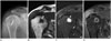
Fig. 2
A 59-year-old woman with an intraosseous lipoma (MRI stage 3) in proximal metaphysis of right femur.
A. Anteroposterior radiograph shows a poorly defined geographic lesion in intertrochanteric portion of the right femur with reticular calcification.
B. On coronal T1-weighted MR image, high signal intensity (arrowheads) in peripheral portion of the lesion demonstrate fat component of the lesion, corresponding to low signal intensity in fat-saturated T2-weighted MR image.
C. Cystic change in the center of the lesion shows high signal intensity on coronal fat-saturated T2-weighted MR image.
D. Contrast enhanced fat-saturated T1-weighted MR image shows subtle rim enhancement of cystic change.
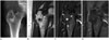
Fig. 3
A 57-year-old man with an intraosseous lipoma (MRI stage 3) in distal metaphysis of right tibia.
A. Anteroposterior radiograph shows an expansive osteolytic lesion with well-defined border, sclerotic rim, and internal ring calcifications.
B. Coronal T1-weighted MR image shows well-defined mass with peripheral high signal intensity and central cystic changes (arrowheads).
C. Coronal fat-saturated T2-weighted MR image shows cystic changes (arrowheads) with high signal intensity. Marginal low signal intensity reveals internal round calcification.
D. Contrast enhanced fat-saturated T1-weighted MR image shows subtle rim enhancement of cystic changes. Curettage and bone cement insertion was conducted to prevent expected osteoarthritis and pathologic fracture. Lesion was confirmed histopathologically.
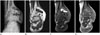
Fig. 4
A 48-year-old man with an intraosseous lipoma (MRI stage 3) in proximal diaphysis of right humerus.
A. Anteroposterior radiograph shows an osteolytic lesion with sclerotic rim.
B. Small fatty portion of intraosseous lipoma (arrowhead) and adjacent bone marrow have similar high signal intensity in T1-weighted MR image. Thin low signal intensity rims demarcate intraosseous lipoma and internal cystic change.
C. It is difficult to differentiate peripheral fatty portion (arrowhead) of intraosseous lipoma with adjacent fatty bone marrow in fat-saturated T2-weighted MR image. This case demonstrates that intraosseous lipoma containing small fat and extensive cystic change can be misdiagnosed as other cystic bone lesion.
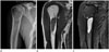
Table 1
Age Distribution of Intraosseous Lipomas in Long Bones
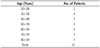
| Age (Years) | No. of Patients |
|---|---|
| 20–29 | 3 |
| 30–39 | 4 |
| 40–49 | 7 |
| 50–59 | 7 |
| 60–69 | 4 |
| 70–79 | 1 |
| 80–89 | 1 |
| Total | 27 |
Table 2
Involved Bones and Sites of Intraosseous Lipomas in Long Bones
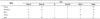
| Bone | Site | |||||
|---|---|---|---|---|---|---|
| Prox E | Prox M | D | Dist M | Dist E | Total | |
| Femur | 1 | 9 | 1 | 0 | 1 | 12 |
| Humerus | 3 | 3 | 1 | 1 | 0 | 8 |
| Tibia | 0 | 1 | 1 | 1 | 1 | 4 |
| Fibula | 0 | 1 | 1 | 1 | 0 | 3 |
| Total | 4 | 14 | 4 | 3 | 2 | 27 |
Table 3
Size of Intraosseous Lipomas in Long Bones
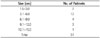
| Size (cm) | No. of Patients |
|---|---|
| 1.5–3.0 | 7 |
| 3.1–6.0 | 12 |
| 6.1–9.0 | 4 |
| 9.1–12.0 | 3 |
| 12.1–15.0 | 1 |
| Total | 27 |
Table 4
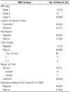
Radiographic Findings of Intraosseous Lipomas in Long Bones (n = 27)
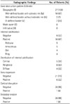
Table 5
MRI Findings of Intraosseous Lipomas in Long Bones (n = 27)
References
1. Reig-Boix V, Guinot-Tormo J, Risent-Martinez F, Aparisi-Rodriguez F, Ferrer-Jimenez R. Computed tomography of intraosseous lipoma of os calcis. Clin Orthop Relat Res. 1987; (221):286–291.
2. Chow LT, Lee KC. Intraosseous lipoma. A clinicopathologic study of nine cases. Am J Surg Pathol. 1992; 16:401–410.
3. Pappas AJ, Haffner KE, Mendicino SS. An intraosseous lipoma of the calcaneus: a case report. J Foot Ankle Surg. 2014; 53:638–642.
4. Bagatur AE, Yalcinkaya M, Dogan A, Gur S, Mumcuoglu E, Albayrak M. Surgery is not always necessary in intraosseous lipoma. Orthopedics. 2010; 05. 12. DOI: 10.3928/01477447-20100329-13. [Epub].
5. Weinfeld GD, Yu GV, Good JJ. Intraosseous lipoma of the calcaneus: a review and report of four cases. J Foot Ankle Surg. 2002; 41:398–411.
6. Milgram JW. Intraosseous lipomas. A clinicopathologic study of 66 cases. Clin Orthop Relat Res. 1988; (231):277–302.
7. Campbell RS, Grainger AJ, Mangham DC, Beggs I, Teh J, Davies AM. Intraosseous lipoma: report of 35 new cases and a review of the literature. Skeletal Radiol. 2003; 32:209–222.
8. Bano S, Yadav SN, Chaudhary V, Jain VK. Radiological evaluation of bilateral intraosseous calcaneal lipoma in various stages of involution. Eur J Radiol Extra. 2011; 78:e57–e59.
9. Radl R, Leithner A, Machacek F, Cetin E, Koehler W, Koppany B, et al. Intraosseous lipoma: retrospective analysis of 29 patients. Int Orthop. 2004; 28:374–378.
10. Muthuphei MN. Intra-osseous lipoma of the calcaneus. S Afr Med J. 1996; 86:1554–1555.
11. Gonzalez JV, Stuck RM, Streit N. Intraosseous lipoma of the calcaneus: a clinicopathologic study of three cases. J Foot Ankle Surg. 1997; 36:306–310. discussion 329.
12. Greenspan A, Raiszadeh K, Riley GM, Matthews D. Intraosseous lipoma of the calcaneus. Foot Ankle Int. 1997; 18:53–56.
13. Tejero A, Arenas AJ, Sola R. Bilateral intraosseous lipoma of the calcaneus. A case report. Acta Orthop Belg. 1999; 65:525–527.
14. Bertram C, Popken F, Rütt J. Intraosseous lipoma of the calcaneus. Langenbecks Arch Surg. 2001; 386:313–317.
15. Yildiz HY, Altinok D, Saglik Y. Bilateral calcaneal intraosseous lipoma: a case report. Foot Ankle Int. 2002; 23:60–63.
16. Jebson PJ, Schock EJ, Biermann JS. Intraosseous lipoma of the proximal radius with extraosseous extension and a secondary posterior interosseous nerve palsy. Am J Orthop (Belle Mead NJ). 2002; 31:413–416.
17. Palczewski P, Swiątkowski J, Gołębiowski M, Błasińska-Przerwa K. Intraosseous lipomas: a report of six cases and a review of literature. Pol J Radiol. 2011; 76:52–59.
18. Rabbani SA, Ilyas I, Alrumaih H. A rare presentation of an intraosseous lipoma in the proximal femur. Am J Case Rep. 2013; 14:362–365.
19. Schatz SG, Dipaola JD, D'Agostino A, Hanna R, Quinn SF. Intraosseous lipoma of the calcaneus. J Foot Surg. 1992; 31:381–384.
20. Shin DS, Kwak ES, Choi JH. Intraosseous Lipoma. J Korean Orthop Assoc. 2003; 38:526–530.
21. Chung CB, Murphey M, Cho G, Schweitzer M, Hodler J, Haghihi P, et al. Osseous lesions of the pelvis and long tubular bones containing both fat and fluid-like signal intensity: an analysis of 28 patients. Eur J Radiol. 2005; 53:103–109.
22. Kamekura S, Nakamura K, Oda H, Inokuchi K, Iijima T, Ishida T. Involuted intraosseous lipoma of the sacrum showing high signal intensity on T1-weighted magnetic resonance imaging (MRI). J Orthop Sci. 2001; 6:183–186.
23. Bruni L. The "cockade" image: a diagnostic sign of calcaneum intraosseous lipoma. Rays. 1986; 11:51–54.
24. Miller TT. Bone tumors and tumorlike conditions: analysis with conventional radiography. Radiology. 2008; 246:662–674.
25. Park YC, Lee YH, Jung KJ, Sung NK, Chung DS, Kim OD, et al. MR Imaging findings of intraosseous lipoma. J Korean Radiol Soc. 2000; 43:343–348.
26. Murphey MD, Carroll JF, Flemming DJ, Pope TL, Gannon FH, Kransdorf MJ. From the archives of the AFIP: benign musculoskeletal lipomatous lesions. Radiographics. 2004; 24:1433–1466.
27. Blacksin MF, Ende N, Benevenia J. Magnetic resonance imaging of intraosseous lipomas: a radiologic-pathologic correlation. Skeletal Radiol. 1995; 24:37–41.
28. Murphey MD, Arcara LK, Fanburg-Smith J. From the archives of the AFIP: imaging of musculoskeletal liposarcoma with radiologic-pathologic correlation. Radiographics. 2005; 25:1371–1395.
29. Öztekin Ö, Argin M, Oktay A, Arkun R. Intraosseous lipoma: radiological findings. Radiol Bras. 2008; 41:81–86.
30. Milgram JW. Malignant transformation in bone lipomas. Skeletal Radiol. 1990; 19:347–352.
 XML Download