

 PDF
PDF ePub
ePub Citation
 Print
Print


Angiosarcomas are the most common sarcomas of the breasts but they are extremely rare, accounting for only 0.04% of primary breast tumors (1). Because of its rarity, this malignant tumor can be easily misdiagnosed; moreover, this tumor has a high mortality rate. Additionally, radiologic findings are typically not specific; therefore, an accurate diagnosis by a radiologist is difficult (234).
A few cases of angiosarcoma of the breast have been reported; however, most reported cases are neither bilateral nor primary (5). We report herein a case of primary bilateral angiosarcoma of the breast in a young woman. She was treated with a right mastectomy and left wide local excision followed by six cycles of adjuvant chemotherapy. However, multiple metastatic lesions were found upon follow-up radiologic evaluation 3 years later.
CASE REPORT
34-year-old woman with a nonspecific medical history incidentally identified a palpable mass in the upper portion of her right breast and visited a medical clinic. She did not complain of tenderness or pain. She was transported to our hospital for removal of angiosarcoma of the right breast, which was diagnosed using a core needle biopsy.
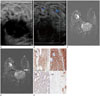
Upon physical examination, a large palpable mass was identified in the upper inner portion of the right breast. The lesion did not show regional skin change. On mammography, both breasts were found to be extremely dense, which lowers the sensitivity of mammography. Real-time gray scale and color Doppler ultrasonography showed a 3.9 × 1.5 × 2.6 cm irregular, circumscribed, hypoechoic mass at the 12 o'clock position in the right breast. The mass was located in the glandular layer, and increased vascularity was not found around or within the mass (Fig. 1A, B). Significantly enlarged lymph nodes were not observed in axillary areas.
Contrast-enhanced breast magnetic resonance imaging (MRI) was performed to evaluate the extent of this mass prior to surgery. A 4.3 × 3.6 × 4.6 cm sized heterogeneously enhancing, irregular mass was found in the right breast (Fig. 1C). The mass showed iso signal intensity compared to the breast parenchyma on T1-weighted images, with high signal intensity on T2-weighted images. Some areas within the mass showed high signal intensity on T1-weighted images and low signal intensity on T2-weighted images. Another 9 × 6 × 7 mm oval, well enhancing mass was detected in the outer portion of the left breast. This mass showed high signal intensity compared to the breast parenchyma on T1- and T2-weighted images (Fig. 1D).
Because the lesion was pathologically diagnosed as an angiosarcoma via a core needle biopsy and radiologic findings were strongly suggestive of malignancy, the attending surgeons performed a right mastectomy and local excision of the mass in the left breast.
Masses from both breasts were pathologically confirmed as angiosarcoma grade III after dissection. A vascular lesion was found in the central portion of the right breast mass. The resection margin was free from carcinoma, and no lymph node metastasis was found. Immunohistochemical staining was positive for CD34, CD31, and actin, but it was negative for the S-100 protein (Fig. 1E). After surgery, a positron emission tomography (PET)-CT scan confirmed that there was no metastatic lesion or residual malignancy.
Because of the high rate of recurrence, the patient was administered six cycles of adjuvant chemotherapy, and radiologic evaluations were performed annually. Three years following the first diagnosis, a metastatic lesion was found in the L1 vertebra via a bone scan and PET-CT. This lesion was treated with additional radiotherapy and chemotherapy; however, a follow-up examination revealed an increase in the size and number of metastatic lesions (in both lungs, the lumbar spine, both clavicles, ribs, iliac bone, and the right pelvic peritoneum).
DISCUSSION
Angiosarcoma of the breast was first described by Schmidt in 1887. This malignant tumor has a vascular endothelial origin and it has a poor prognosis because of high rates of metastasis and local recurrence (6). The prognosis of angiosarcoma depends on its histological grade: low-grade angiosarcoma shows a better survival rate than high-grade angiosarcoma. The 5-year survival rate is 76% for low-grade, 70% for intermediate, and 15% for high-grade angiosarcoma (7).
There are two types of angiosarcoma of the breast: primary and secondary. Secondary angiosarcoma is more common and it typically arises in an irradiated breast after breast conservation or lymphedema, mostly in postmenopausal patients. Primary angiosarcoma is rarer and it tends to sporadically arise in younger women; the median age at diagnosis is the third and fourth decades of life (1). Risk factors for primary angiosarcoma are not known.
Angiosarcomas arise at a young age and display a rapid growth pattern, and most patients identify an abnormality in their breast before a screening mammogram. Occasionally, discoloration of the skin may accompany the lesion because of vascularization (8). In the present case, the patient was a premenopausal 34-year-old woman, had no significant medical history, and presented with a palpable mass in a single breast without skin color change.
A radiologic diagnosis of primary angiosarcoma is difficult due to the young patient population and nonspecific findings. Mammography for angiosarcoma shows typical circumscribed or obscured round, oval, lobulated margins (4910). Additionally, because patients with angiosarcoma are young and typically have dense breasts, a mammogram is not appropriate for identifying this tumor. In our case, mammography also revealed dense breasts. Ultrasonography can provide a wide spectrum of findings: ultrasonography may show heterogeneous hypoechoic or hyperechoic masses with or without posterior acoustic shadowing. However, hypervascularity is usually present, which is in contrast to our case (4910). Thus, owing to these challenges in radiologic diagnosis of angiosarcoma, MRI is thought to be the best modality for diagnosing angiosarcoma.
Many cases of angiosarcoma of the breast show low signal intensity on T1-weighted images and high signal intensity on T2-weighted images as well as early enhancement with prolongation on a dynamic study (3). In our case, the tumors were found to show iso- to high signal intensity on T1-weighted images, high signal intensity on T2-weighted images, and early enhancement with prolongation on a dynamic study. Only the enhancing pattern was similar to the previously reported findings.
The diagnosis of angiosarcoma of the breast is challenging for the radiologist. The young patient population, nonspecific radiological findings, and ordinary symptoms present challenges for the radiologist. Dynamic enhancing MRI and histologic examination should be performed for a suspected angiosarcoma of the breast.
 XML Download