

 PDF
PDF ePub
ePub Citation
 Print
Print


Pancreatic arteriovenous malformation (AVM) is a rare condition defined as a tumorous formation or vascular anomaly arising from an aberrant bypass anastomosis of the arterial and venous systems in the pancreas (1). Esophageal varix bleeding by portal venous hypertension is the most common complication of pancreatic AVM. Duodenal ulcer bleeding or bleeding from the pancreatic AVM itself are other causes of bleeding associated with this disease entity. Pancreatic AVM presenting as retroperitoneal bleeding is an extremely rare clinical condition and is often misdiagnosed as a pseudoaneurysm. Treatment may include ligation of the afferent artery, embolization of feeding vessels, portacaval shunting, or surgical resection of the affected part or the entire affected organ (2).
We report a case of pancreatic AVM presenting as a mesenteric hemorrhage that was successfully treated with transcatheter arterial embolization using n-butyl-2-cyanoacrylate (NBCA).
CASE REPORT
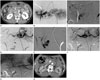
A 62-year-old man presented to the emergency department of our hospital with acute abdominal pain. He had no medical history of alcohol abuse and no history of trauma. He experienced sudden-onset of severe periumbilical pain during defecation the morning on the day of admission. His vital signs were stable. Physical examination revealed mild periumbilical and rebound tenderness. Laboratory data showed a mild increase in white blood cell count (11600/µL) and increased lactate dehydrogenase level (938 IU/L). His hemoglobin, alanine aminotransferase, aspartate aminotransferase, amylase, and lipase levels were normal. A dynamic abdominal computed tomography (CT) scan showed diffuse hemorrhagic infiltration of the mesenteric root and peripancreatic area with compression of the superior mesenteric vein. In addition, a 1-cm nodular enhancing lesion was found adjacent to the superior mesenteric artery (SMA) and dorsomedial to the pancreatic head, which was prospectively diagnosed as a pseudoaneurysm of the mesenteric artery with mesenteric and peripancreatic hemorrhages (Fig. 1A). He was referred to the intervention department for further evaluation. Selective superior mesenteric arteriography revealed a nodular nidus at the pancreatic head that was fed by a small branch of the inferior pancreaticoduodenal artery with a drainage vein to the upper superior mesenteric vein and early visualization of the portal vein (Fig. 1B, C). Another feeder of AVM was the dorsal pancreatic artery, branching from the proximal common hepatic artery just distal to the splenic artery bifurcation on celiac arteriogram (Fig. 1D, E). These findings were consistent with a pancreatic AVM. Owing to the relatively large extent of the hemorrhage and the presence of only two feeding arteries, endovascular treatment was considered to avoid invasive surgical excision. The feeding artery via the small branch of the inferior pancreaticoduodenal artery was easily super selected with a 2.4-Fr microcatheter (Progreat, Terumo, Tokyo, Japan) through the SMA approach. However, the other feeding artery via the dorsal pancreatic artery was successfully catheterized retrogradely through the pancreaticoduodenal arcade, after a failed attempt through the celiac approach due to the very acute origin of the target artery. The feeding arteries were embolized with injection of NBCA (Histoacryl, B. Braun, Melsungen, Germany) diluted 1:4 (first artery) and 1:2 (second artery) with iodized oil (Lipiodol, Guerbet, Aulnay-Sous-Bois, France). A post-procedure angiography revealed complete exclusion of the feeding arteries, nidus, and aberrant draining vein (Fig. 1F, G). The post-procedure course was uneventful, and the patient's symptoms abated after the procedure. A follow-up CT scan obtained 2 months after the procedure showed complete disappearance of the pancreatic AVM and mesenteric hemorrhage (Fig. 1H).
DISCUSSION
Pancreatic AVM is a rare condition defined as a tumorous formation or vascular anomaly arising from an aberrant bypass anastomosis of the arterial and venous systems in the pancreas (1). Halpern et al. (1) first reported a patient with pancreatic AVM associated with Rendu-Osler-Weber disease in 1968. Pancreatic AVM is classified as a congenital anomaly thought to be caused by failure of the regulatory sphincteric mechanism of the arteriole-capillary junction, resulting in unrestricted overflow of arterial blood into the capillary bed, or by acquired disease complicated by trauma, inflammation, or tumor.
Pancreatic AVM has various clinical manifestations, which may be asymptomatic or include abdominal pain, pancreatitis, and/or gastrointestinal (GI) bleeding. Nishiyama et al. (2) reviewed the cases of 42 patients with pancreatic AVM. They reported that 50% of patients presented with life-threatening GI bleeding. The AVM was located in the pancreatic head in 56% of the patients, 31% of whom also had an extrapancreatic AVM (2). Makhoul et al. (3) classified the bleeding mechanisms of pancreatic AVM into two categories as follows: first, bleeding from a ruptured esophageal or gastric varix secondary to portal hypertension induced by the AVM; and second, hemorrhage from direct erosion of the AVM into the pancreatic duct or through the adjacent intestinal mucosa as a duodenal ulcer. However, pancreatic AVM presenting as retroperitoneal bleeding has been rarely documented. In the present case, the hemorrhage was present in the small bowel mesentery and peripancreatic retroperitoneal space. As the patient's pain developed acutely during defecation, we assume that this rare instance of retroperitoneal bleeding was the result of an abrupt increase in portal venous pressure induced by increased intraperitoneal pressure.
Doppler ultrasonography is a non-invasive and easily repeated modality that can be performed at a patient's bedside. Pancreatic AVM appears on Doppler ultrasonography as mosaic lesions composed of pulsatile waves, but ultrasonography has limitations in revealing the full extent of the lesion and its relation to adjacent organs. The characteristic CT findings of AVM include nod-ular enhancement and early appearance of the portal venous system. Although nodular enhancement and early appearance of the portal venous system were observed in 76.9% and 66.7% of patients, respectively, in one study (4), some cases demonstrated uncharacteristic imaging findings that led to misdiagnosis as a pseudoaneurysm or a true aneurysm, as in the present case. Magnetic resonance imaging (MRI) shows a tangle of tubular structures with a signal void on T2-weighted image for high blood flow and hyperintense enhancement of tubular structures on enhanced arterial phase, as a characteristic feature of AVM (5). However, MRI is not a widely used imaging modality for cases of acute bleeding. Dilated and tortuous feeding arteries, racemose staining in the nidus, and early appearance of the portal venous system have been reported as characteristic angiographic findings of AVM (4).
The treatment methods for pancreatic AVM are surgery (57.1%); nonsurgical therapy, including embolization, irradiation, or a portovenous shunt (14.3%); and no treatment (28.6%) (2). Surgery is the least preferable treatment method because total extirpation of the affected organ, or at least the involved por-tion, is the only way to assure a complete cure and eliminate the possibility of recurrent bleeding. However, excision of the pancreatic head is a highly invasive procedure that carries a high risk of complications. Thus, the efficacy of surgery should be determined after considering the size and location of the lesion. Surgery has also been reported to possibly have a limited value because of inoperability or inaccessibility of the affected region (2). An endovascular approach is a good alternative for poor surgical candidates. Transarterial embolization (TAE) has been shown to be a good alternative treatment for the control of hemorrhage from various bleeding foci in cases of congenital AVM (6). Tasuta et al. (7) reported successful treatment of cases with TAE alone in patients without hemorrhagic complications. In their summary of 13 previous reports of pancreatic AVM treated with TAE, all patients with hemorrhagic complication initially treated with TAE required surgery. The recurrent bleeding rate after successful TAE has been reported to be up to 37% (7). Although complete regression of pancreatic AVM with numerous feeding arteries is difficult to achieve using TAE; complete embolization of the feeding arteries, nidus, and draining veins is achievable in patients with a considerable number of feeding arteries. Successful treatment of pancreatic AVM using TAE with NBCA, or ethylene vinyl alcohol copolymer (Onyx), has been reported (78). Moreover, a transvenous approach has been reported to be a useful endovascular approach in patients with too many collateral arteries to embolize separately (6). However, transvenous embolization carries a high risk of increasing venous pressure, resulting in catastrophic bleeding from the AVM. The commonly used embolic agents are metallic coil, gelatin sponge, and poly-vinyl-alcohol (PVA) particles. Metallic coil is an effective embolic agent, but it can embolize only the feeding artery, excluding the nidus and draining vein. Thus, the recurrence rate could be high through multiple other feeders. Furthermore, gelatin slurry and PVA particles carries the risk of migration of considerable amount of embolic agent to the distal vein via AVM, until complete occlusion is achieved. NBCA or Onyx is a commonly used permanent liquid embolic agent in TAE for AVMs in various other organs. NBCA and Onyx can achieve not only embolization of the feeding artery but also distal vasculature, including the nidus and draining vein. In the present case, only two feeding arteries to the pancreatic AVM were found on arteriography of the SMA and celiac artery. Super selection of the two feeders was successfully achieved using a microcatheter. For flow occlusion, we used a mixture of NBCA and iodized oil at ratios of 1:2 and 1:4 as a permanent embolization agent to delay polymerization for complete embolization of not only the feeding arteries but also the draining veins. No considerable distal migration of NBCA to the portal vein was observed. Although low concentration of NBCA injection may result in reflux or distal migration of NBCA, which can lead to non-target embolization, a small amount of NBCA may not influence the main portal flow or liver function. The NBCA concentration should be adequately adjusted for its polymerization time to embolize the draining vein.
The present patient was followed up for only 2 months, but complete exclusion of the AVM with the disappearance of the nidus and the draining vein was observed on the follow-up CT scan.
In conclusion, we report a rare case of pancreatic AVM presenting as retroperitoneal bleeding, which was successfully treated with TAE without any complications.
 XML Download