

 PDF
PDF ePub
ePub Citation
 Print
Print


INTRODUCTION
While uncommon, pulmonary artery aneurysms and pseudoaneurysms are associated with high mortality. Overall they are considered rare entities with prevalence rates of around 1 case per 14000 to 100000 autopsies (12). Most pulmonary artery aneurysms and pseudoaneurysms are acquired and associated with cardiovascular disease, infection, iatrogenic causes, trauma, neoplasm, and connective tissue disease. To our knowledge, while there are many cases of Rasmussen aneurysm in patients with cavitary tuberculosis, there have been no reports of pseudoaneurysm due to fungus ball in the progressive massive fibrosis (PMF) of a patient with pneumoconiosis. We described a case of pseudoaneurysm detected by contrast-enhanced chest CT with three-dimensional (3D) reconstruction. The pseudoaneurysm arose in the upper lobar branch of the right pulmonary artery and caused by a fungus ball within the PMF in a patient with pneumoconiosis, who underwent transcatheter endovascular embolization of the aneurysm.
CASE REPORT
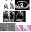
The patient was a 51-year-old man with a history of pneumoconiosis, tuberculosis, and hypertension. He was a coal miner for 12 years and had a 30 pack-year smoking history. He initially presented to another hospital with an episode of pneumonia and was transferred to our hospital for recurrent episodes of massive hemoptysis of more than 1 liter per day. On hospital admission, the patient was afebrile and hemodynamically stable (134/85 mm Hg, 106 beats per minute, 20 breaths per minute, and temperature of 36.8℃). The initial laboratory results showed anemia, mild leukocytosis (hemoglobin: 7.6 g/dL, hematocrit: 22.7%, and white blood cell count: 10300/µL) and negative three sputum acid fast bacillus smears and culture. Contrast-enhanced chest CT with 3D reconstruction (LightSpeed 16; GE Medical Systems, Milwaukee, WI, USA) showed multiple perilymphangitic nodules suggestive of pneumoconiosis (Fig. 1A), spongiform air bubbles indicating a fungus ball within a PMF, and a tuberculous scar with volume loss and traction bronchiectasis. There was no three-in-bud or centrilobular nodule, cavitary nodule, lymphadenopathy, or effusion that was suggestive of active tuberculosis. Within the fungus ball, a pseudoaneurysm was noted in the upper lobar branch of the right pulmonary artery (Fig. 1B-D). The lack of a draining vein ruled out an arteriovenous malformation. Emergent pulmonary artery angiogram and transcatheter embolization were performed with 4–5 mm coils (Tornado; Cook, Bloomington, IN, USA) via a right femoral venous approach (Fig. 1E, F). Particle embolization (Contour; Boston Scientific, Natick, MA, USA) of the right intercostobronchial trunk and right 1st-2nd intercostal artery arising from the costocervical trunk were also performed due to arterial hypertrophy. Following these procedures, the hemoptysis stopped, but recurred at not more than 20 cc per day after one week. Because of recurrent hemoptysis and deteriorating pulmonary function test results, a right upper lobe lobectomy was performed for the pulmonary aspergilloma with pulmonary artery pseudoaneurysm. Although the presence of cavitation in a conglomerate mass is an important indication of associated tuberculosis, the pathologic specimen of this case showed fungal hyphae within the PMF cavity without chronic granulomatous inflammation suggesting tuberculosis (Fig. 1G, H). Polarized light images showed scattered interstitial silica particles. The postoperative period was uneventful and the patient was discharged in the fourth postoperative week.
DISCUSSION
Pulmonary artery aneurysms and pseudoaneurysms can be congenital or acquired. Common congenital causes include vessel wall deficiency, valvular and postvalvular stenosis, and increased flow due to left-to-right shunts. Common acquired causes include trauma, pulmonary artery hypertension, vasculitis (Behçet's syndrome and Hughes-Stovin syndrome), infection (mycotic aneurysms and pseudoaneurysms), neoplasm, iatrogenic causes (malpositioned Swan-Ganz catheters), and connective tissue abnormalities such as Marfan syndrome, Ehlers-Danlos syndrome, and cystic medial necrosis (34). There have been many reports of pulmonary artery pseudoaneurysm due to tuberculosis or fungus infection. Also, fungus infection is well known in patients with pneumoconiosis. To the best of our knowledge, there have been no reports of pseudoaneurysm due to fungus ball in the PMF of a patient with pneumoconiosis. Therefore, we reported the first case of pseudoaneurysm due to fungus ball in the PMF of a patient with pneumoconiosis.
The cavity formation within the PMF suggests ischemic necrosis due to deficient blood supply (5). PMF greater than 4 cm may show cavitation due to ischemic necrosis. Tuberculosis is another possible cause of cavitation in PMF. The presence of cavitation in a conglomerate mass (PMF) is an important indication of associated tuberculosis (6). Kato et al. (7) proposed that chronic persistent or progressive upper lobe infiltrates and cavities in patients with pneumoconiosis should raise the possibility of chronic necrotizing pulmonary aspergillosis. Preexisting diseases associated with aspergilloma include tuberculosis, bronchiectasis, pneumoconiosis, and sarcoidosis (8). Aspergillomas occur predominantly in the upper lobes, reflecting a predilection for cavity formation at this site, because the relative imbalance in perfusion and ventilation in the lung apices provide an oxygen-rich environment for micro-organisms (9).
Our case showed spongiform air bubbles in the PMF cavity suggestive of fungus ball. Ischemic necrosis of PMF due to deficient blood supply might cause cavitation of PMF and subsequent fungal hyphae growth in the PMF cavity. Mycotic pulmonary artery pseudoaneurysms are caused by several mechanisms such as direct extension of inflammation to involve the vessel wall, endovascular seeding of the vessel wall from bronchial arteries in septicemia and intimal invasion of the vessel wall from septic embolism. In our case, it is likely that the fungal hyphae eroded and weakened the branch of the pulmonary artery, leading to the vessel pseudoaneurysm.
CT is useful for diagnosing diseases that cause massive hemoptysis, localizing bleeding sites, and selecting vessels to be embolized (10). Contrast-enhanced chest CT with 3D reconstruction scan helped us to detect pseudoaneurysm of the pulmonary artery in PMF, because the enhancement density of the pseudoaneurysm of pulmonary artery was similar to that of the aorta. After detecting the cause of massive hemoptysis such as pseudoaneurysm, we suggest that embolization of pseudoaneurysm due to fungus ball within the PMF is necessary to stop abrupt massive pulmonary bleeding. After that, surgical removal of PMF with fungus ball is the best way to prevent recurrent hemoptysis.
In conclusion, we reported a case of pseudoaneurysm due to fungus ball in the PMF of a patient with pneumoconiosis. Assessment with contrast-enhanced CT with coronal reconstruction images allowed accurate evaluation of pulmonary artery pseudoaneurysm, facilitating prompt diagnosis and treatment.
 XML Download