

 PDF
PDF ePub
ePub Citation
 Print
Print


INTRODUCTION
An intercoronary communication is a direct communication between the two main coronary arteries and it can develop in the absence of an obstructive lesion (12). Its true prevalence in the general population is not known, but the documented coronary angiographic incidence is 0.002% in 126595 patients and 0.02% in 9726 patients (13). It can be distinguished from collateral arteries by its angiographic features, and it does not reflect an underlying coronary disease (2). Here, we present a case of intercoronary communication between the right coronary artery (RCA) and the left circumflex coronary artery (LCX) found incidentally in a 48-year-old man. The initial diagnosis was made using electrocardiogram (ECG)-gated multidetector computed tomography (MDCT).
CASE REPORT
A 48-year-old man presented to the hospital with chest tightness and acute exertional chest pain. His symptoms had developed 5 days earlier. There was no history of trauma, hypertension, diabetes, or surgery. He did not have any connective tissue disorder or any other systemic anomaly, and there was no notable family history of disease. The physical examination was normal, with an arterial blood pressure of 130/90 mm Hg. A 12-lead ECG showed a normal sinus rhythm with a heart rate of 65 beats/min. Chest radiography revealed a normal-sized heart contour with no lung infiltration. Transthoracic echocardiography showed normal-sized cardiac chambers and normal left ventricular systolic function.
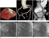
Cardiac CT performed using a 128-slice MDCT scanner (Ingenuity Core 128; Philips Medical Systems, Best, the Netherlands) showed no significant coronary artery stenosis, pulmonary thromboembolism and aortic dissection. However, an intercoronary communication between the posterolateral branch of the RCA and the distal LCX was noted (Fig. 1A-C, Supplementary Movie 1 in the online-only Data Supplement). Coronary angiography revealed no significant luminal narrowing or occlusion of the coronary arteries. However, selective and simultaneous injection into the right and left coronary ostia confirmed the interarterial continuity between the RCA and LCX (Fig. 1D-F, Supplementary Movie 2 in the online-only Data Supplement). Selective injection into the left coronary artery showed retrograde filling of the RCA from the distal LCX, while right coronary injection filled the RCA and LCX. Retrograde filling did not involve the collaterals, but it was rather due to a bidirectional intercoronary communication. The patient was reassured that his coronary arteries were normal. Medical treatment (aspirin 100 mg/day, metoprolol 50 mg/day, and isosorbide mononitrate 40 mg/day) alone controlled his symptoms, and he was discharged in good condition.
DISCUSSION
Intercoronary arterial communication or "coronary arcade" is a rare variant of the coronary circulation (12). It is distinguished from the coronary collaterals seen in patients with occlusive coronary artery disease based on the angiographic features and histological structure (4). In the presence of occlusive coronary artery disease, collaterals dilate progressively in an attempt to mitigate the severity of myocardial ischemia and necrosis. In comparison, intercoronary communications are single, extramural, straight or gently curved, and larger in diameter (> 1 mm) than collaterals. They also appear to be histologically different: collaterals that develop in the presence of obstructive coronary artery disease are composed of endothelium supported by poorly organized collagen, muscle, and elastic fibers, whereas intercoronary communications are similar to an epicardial vessel with a well-defined muscle layer (45). Rare cases of intercoronary communication in the absence of obstructive coronary artery disease have been reported (123456). In the past, intercoronary communications were usually diagnosed postmortem or by cardiac catheterization. Conventional coronary angiography is the gold standard for assessing coronary arteries. However, recent advances in cardiac MDCT enable non-invasive diagnosis of this condition and other coronary anomalies (78). In our patient, ECG-gated, cardiac MDCT was performed for triple rule-out (acute coronary syndrome, pulmonary thromboembolism, and aortic dissection) and an intercoronary communication between the posterolateral branch of the RCA and the distal LCX was discovered incidentally by MDCT.
The functional significance of intercoronary communications is unclear. They may have a protective role if occlusive coronary lesions develop in any of the involved vessels. However, Donaldson and Isner (5) reported a case with > 95% narrowing of the RCA, in which the left anterior descending artery–posterior descending artery (PDA) continuity did not prevent extensive transmural infarction in the distribution of the PDA; hence, the theoretical protective role is questionable. Furthermore, myocardial ischemia can result from a coronary steal by the unidirectional intercoronary communication (26). Kim et al. (4) and Takatsu et al. (9) reported cases of intercoronary communications with vasospastic angina. Although the relation between intercoronary communication and coronary artery spasm is not clear, consideration of coronary vasospasm can be useful if intercoronary communication without significant coronary stenosis is found in patients with chest pain. Fortunately, our patient did not have combined significant coronary artery stenosis or any other disease; medical treatment (aspirin, metoprolol, and isosorbide mononitrate) controlled his symptoms. Further studies are needed to evaluate the potential role of intercoronary communication and to establish a standard management protocol.
In summary, we report a case of intercoronary communication between the RCA and LCX found incidentally in a 48-year-old man who was initially diagnosed using ECG-gated MDCT.
SUPPLEMENTARY MOVIE LEGENDS
Communication between the right and circumflex coronary arteries discovered incidentally by MDCT.
Movie 1. Maximum intensity projection cardiac MDCT axial images during mid-diastole.
Movie 2. Coronary angiographic images in the left anterior oblique caudal view with simultaneous injection of contrast material into both coronary ostia.
 XML Download