

 PDF
PDF ePub
ePub Citation
 Print
Print


Abstract
Varicocele is a dilatation of the veins in the pampiniform plexus and manifests as mass-effect, pain, testicular atrophy, or male infertility. Traditionally, surgical treatment has been the mainstay of treatment of varicocele, while interventional treatment, which is endovascular embolization of the testicular vein, has been gaining popularity recently. In this review, diagnosis of the disease, indications and procedure details of interventional treatment, results, and complications are discussed.
Figures and Tables
Fig. 1
Ultrasonographic features of a varicocele.
A. On ultrasonographic image, pampiniform plexus of veins is dilated more than 2 mm in diameter.
B. Color Doppler image shows hypervascularity of the dilated pampiniform plexus.
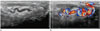
Fig. 3
Type III varicocele.
Left renal venogram shows reflux of contrast medium into the testicular vein (arrows) (A). The catheter is inserted from above, through the right basilic vein access. The testicular vein shows duplication (arrows) in its distal part (B). Note the contrast medium reflux into the pampiniform plexus (arrowhead) (B). The distal testicular vein is embolized with coils and treated with sclerotherapy (C); then, the proximal testicular vein is also embolized with coils (D). Left renal venogram shows no further reflux into the testicular vein (D).
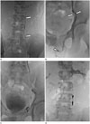
Fig. 4
Type IVb varicocele.
A. Left renal venogram shows reflux of contrast medium into the tortuous collateral vein (arrows). The proximal testicular vein is not seen.
B. The collateral vein (arrows) is connected with the probable testicular vein (arrowheads).
C. The testicular vein (arrows) is refluxed distally below the inguinal canal.
D. The distal testicular vein is embolized with coils and treated with sclerotherapy, followed by coil embolization of the proximal testicular vein.
E. Ultrasonographic image shows a dramatic reduction in the vascularity of the pampiniform plexus six days after sclerotherapy.
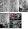
Table 1
Venography of a Left Varicocele (Bähren Classification)
References
1. Shiraishi K, Matsuyama H, Takihara H. Pathophysiology of varicocele in male infertility in the era of assisted reproductive technology. Int J Urol. 2012; 19:538–550.
2. Raheem OA. Surgical management of adolescent varicocele: systematic review of the world literature. Urol Ann. 2013; 5:133–139.
3. Halpern J, Mittal S, Pereira K, Bhatia S, Ramasamy R. Percutaneous embolization of varicocele: technique, indications, relative contraindications, and complications. Asian J Androl. 2016; 18:234–238.
4. Mohammadi A, Ghasemi-Rad M, Mladkova N, Masudi S. Varicocele and nutcracker syndrome: sonographic findings. J Ultrasound Med. 2010; 29:1153–1160.
5. Wright EJ, Young GP, Goldstein M. Reduction in testicular temperature after varicocelectomy in infertile men. Urology. 1997; 50:257–259.
6. Gat Y, Gornish M, Chakraborty J, Perlow A, Levinger U, Pasqualotto F. Azoospermia and maturation arrest: malfunction of valves in erect poster of humans leads to hypoxia in sperm production site. Andrologia. 2010; 42:389–394.
7. Dubin L, Amelar RD. Varicocele size and results of varicocelectomy in selected subfertile men with varicocele. Fertil Steril. 1970; 21:606–609.
8. Mihmanli I, Kurugoglu S, Cantasdemir M, Zulfikar Z, Halit Yilmaz M, Numan F. Color Doppler ultrasound in subclinical varicocele: an attempt to determine new criteria. Eur J Ultrasound. 2000; 12:43–48.
9. Bähren W, Lenz M, Porst H, Wierschin W. [Side effects, complications and contraindications for percutaneous sclerotherapy of the internal spermatic vein in the treatment of idiopathic varicocele]. Rofo. 1983; 138:172–179.
10. Sigmund G, Bähren W, Gall H, Lenz M, Thon W. Idiopathic varicoceles: feasibility of percutaneous sclerotherapy. Radiology. 1987; 164:161–168.
11. Jargiello T, Drelich-Zbroja A, Falkowski A, Sojka M, Pyra K, Szczerbo-Trojanowska M. Endovascular transcatheter embolization of recurrent postsurgical varicocele: anatomic reasons for surgical failure. Acta Radiol. 2015; 56:63–69.
12. Nagappan P, Keene D, Ferrara F, Shabani A, Cervellione RM. Antegrade venography identifies parallel venous duplications in the majority of adolescents with varicocele. J Urol. 2015; 193:286–290.
13. Iaccarino V, Venetucci P. Interventional radiology of male varicocele: current status. Cardiovasc Intervent Radiol. 2012; 35:1263–1280.
14. Kim J, Shin JH, Yoon HK, Ko GY, Gwon DI, Kim EY, et al. Persistent or recurrent varicocoele after failed varicocoelectomy: outcome in patients treated using percutaneous transcatheter embolization. Clin Radiol. 2012; 67:359–365.
15. Storm DW, Hogan MJ, Jayanthi VR. Initial experience with percutaneous selective embolization: a truly minimally invasive treatment of the adolescent varicocele with no risk of hydrocele development. J Pediatr Urol. 2010; 6:567–571.
16. Marsman JW. The aberrantly fed varicocele: frequency, venographic appearance, and results of transcatheter embolization. AJR Am J Roentgenol. 1995; 164:649–657.
17. Gandini R, Konda D, Reale CA, Pampana E, Maresca L, Spinelli A, et al. Male varicocele: transcatheter foam sclerotherapy with sodium tetradecyl sulfate--outcome in 244 patients. Radiology. 2008; 246:612–618.
18. Urbano J, Cabrera M, Alonso-Burgos A. Sclerosis and varicocele embolization with N-butyl cyanoacrylate: experience in 41 patients. Acta Radiol. 2014; 55:179–185.
19. Hawkins CM, Racadio JM, McKinney DN, Racadio JM, Vu DN. Varicocele retrograde embolization with boiling contrast medium and gelatin sponges in adolescent subjects: a clinically effective therapeutic alternative. J Vasc Interv Radiol. 2012; 23:206–210.
20. Li L, Zeng XQ, Li YH. Safety and effectiveness of transcatheter foam sclerotherapy for testicular varicocele with a fluoroscopic tracing technique. J Vasc Interv Radiol. 2010; 21:824–828.
21. Lenz M, Hof N, Kersting-Sommerhoff B, Bautz W. Anatomic variants of the spermatic vein: importance for percutaneous sclerotherapy of idiopathic varicocele. Radiology. 1996; 198:425–431.
22. Wunsch R, Efinger K. The interventional therapy of varicoceles amongst children, adolescents and young men. Eur J Radiol. 2005; 53:46–56.
23. Reiner E, Pollak JS, Henderson KJ, Weiss RM, White RI Jr. Initial experience with 3% sodium tetradecyl sulfate foam and fibered coils for management of adolescent varicocele. J Vasc Interv Radiol. 2008; 19(2 Pt 1):207–210.
24. Sze DY, Kao JS, Frisoli JK, McCallum SW, Kennedy WA 2nd, Razavi MK. Persistent and recurrent postsurgical varicoceles: venographic anatomy and treatment with N-butyl cyanoacrylate embolization. J Vasc Interv Radiol. 2008; 19:539–545.
25. Abdulmaaboud MR, Shokeir AA, Farage Y, Abd El-Rahman A, El-Rakhawy MM, Mutabagani H. Treatment of varicocele: a comparative study of conventional open surgery, percutaneous retrograde sclerotherapy, and laparoscopy. Urology. 1998; 52:294–300.
 XML Download