

 PDF
PDF ePub
ePub Citation
 Print
Print


INTRODUCTION
Leiomyoma is a benign tumor of smooth muscle and can occur in any organ; but the most common form occurs in the uterus, small bowel and esophagus. Involvement of the mediastinum as the primary site of origin is extremely uncommon, with only 14 cases reported in the English literature (12). The most common radiographic finding of primary mediastinal leiomyoma is a well circumscribed heterogeneous solid mass. We reported a case of primary mediastinal leiomyoma that was demonstrated by computed tomography (CT) scan as homogenous hypoattenuating mass, mimicking a giant cyst-like lesion. To our knowledge, this is the first reported case of primary mediastinal leiomyoma presenting with this radiologic finding.
CASE REPORT
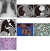
A 58-year-old man was admitted to a local community hospital due to falling from 2 stairs height 8 days prior. He was transferred to our hospital with abnormal findings on chest CT. He had no clinical sign or symptom. Physical and systemic examinations were normal. Laboratory findings showed no abnormalities except for mildly elevated total bilirubin (1.6 mg/dL). Posteroanterior chest radiograph was taken and demonstrated a large left retrocardiac soft tissue mass (Fig. 1A). Contrast enhanced CT scan was obtained with a 128-detector CT scanner (SOMATOM Definition A+ 128, Siemens Medical Solutions, Erlangen, Germany), using 120-kVp technique. Total of 80 mL of intravenous contrast material (300 mg/mL, 1.2 mL/kg) was injected with flow rate of 3.0 mL/s. Automatic trigger technology was used, while the threshold of the main pulmonary artery trunk was set to 141 Hounsfield unit (HU). Contrast enhanced CT images showed a smooth margin, thin-walled, homogenous, hypoattenuated mass of about 112 × 90 mm in the left posterior mediastinum. Mediastinal mass showed about 10-30 HU (pre-contrast CT) (Fig. 1B) and 10-30 HU (post-contrast CT) on region of interest in axial slices (Fig. 1C), which mimics a huge mediastinal cyst or cyst-like lesion that shows almost no enhancement. The mass was closely in contact with the esophagus with suspected esophageal invasion. Smaller bronchi in left lower lobe were upward laterally displaced by the mass (Fig. 1D). Clinical and radiographic differential diagnosis included congenital mediastinal cysts such as bronchogenic cyst, esophageal duplication cyst, neurenteric cyst and cystic lymphangioma. The patient wanted to confirm the mediastinal mass and chose surgery rather than biopsy due to suspicious esophageal invasion. The operative findings showed a huge, well-encapsulated tumor with a solid portion in the middle mediastinum and pleuritic adhesions. However, the tumor showed no definite invasion into the adjacent mediastinal structure. Tumor was resected without complication. The patient had unremarkable postoperative course and was discharged on 11th postoperative days. On gross examination, the resected gross specimen showed softly flesh and mucoid cut surface with hemorrhage in the inner wall (Fig. 1E). Microscopically, there were absence of mitotic count and sparse cellularity. In the immunohistochemical examinations of the excised mass, desmin, smooth muscle actin (SMA), and melanosome-associated protein (HMB-45) were positive; however, D2-40, CD 34, and C-kit were negative (Fig. 1F). The light microscopic features of the tumor showed microcystic changes between slender neoplastic tumor cell clusters (Fig. 1G). Histologic examination of the tumor represented benign leiomyoma with microcystic change.
DISCUSSION
Mediastinal leiomyomas are extremely rare tumors that usually originate from the adjacent structures containing smooth muscle in their wall (esophagus and large vessels) (2). In our case, primary mediastinal leiomyoma showed no tumor dependency to neighboring structures. To our knowledge, only 14 cases of primary mediastinal leiomyoma have been described in the English literature (2). In the study by Suzuki et al. (3), primary mediastinal leiomyoma are most frequently found in middle age group and develop more frequently in women; and the posterior mediastinum is the most common location (4). They are slow-growing with no characteristic symptoms (56).
According to published case reports, CT findings of primary mediastinal leiomyoma are well defined as heterogeneous enhancing solid mass, similar to that of uterine myoma (12). In our case, contrast enhanced CT scan showed a homogenous, low-attenuated, well defined mass. To our knowledge, this is the first reported case of primary mediastinal leiomyoma presenting with this radiologic finding. On histology, microcystic changes within neoplastic tumor cell clusters of the leiomyoma and their mucin production were seen, corresponding to homogenous low attenuating mass.
Differential diagnosis for homogenous low attenuating mass in the middle mediastinum include congenital cysts such as bronchogenic cyst, esophageal duplication cyst, neurentic cyst, cystic lymphangioma, or other cyst-like mass. It is challenging to differentiate homogenous low attenuating masses from true cystic lesions in mediastinum, preoperatively. Esophageal leiomyoma are also seen as incidental homogenous low attenuating mediastinal mass (7). Although it is the most common benign tumor of esophagus, esophageal leiomyoma has only been found in 0.1-0.006% of autopsies (8). CT scan findings of huge esophageal leiomyoma can appear similar to those of our case; so it is difficult to differentiate primary mediastinal leiomyoma from esophageal leiomyoma. In our case, primary mediastinal leiomyoma appeared as a large encapsulated mass that was separated from esophagus.
Surgical resection for definitive diagnosis is the recommended treatment for all suspected leiomyoma of mediastinum whether symptomatic or asymptomatic. The type of operation depends on the location and size of tumor.
The definitive diagnosis of primary mediastinal leiomyoma is achieved by histology. Microscopically, leiomyoma consists of monomorphic spindle cells with blunt-ended nuclei, arranged in interlacing fascicles. The specific immune marker, to SMA, is a diagnostic sign for leiomyoma (2).
The lesion must be distinguished from leiomyosarcoma. In contrast to leiomyosarcoma, histologic features of leiomyoma include absence of mitotic count, low cellularity, lack of cytologic atypia and pleomorphism, and prominent fibrosis or hyalinization. Prognosis for leiomyoma is generally excellent (9).
In conclusion, primary mediastinal leiomyoma is an extremely rare tumor. This is the first report of primary mediastinal leiomyoma that was demonstrated by CT scan as homogenous, hypoattenuating mass without gross enhancement, mimicking a giant cyst like lesion. Thus, primary mediastinal leiomyoma should be considered in differential diagnosis of a huge cyst-like lesion in mediastinum.
 XML Download