

 PDF
PDF ePub
ePub Citation
 Print
Print


INTRODUCTION
In the Republic of Korea, the incidence of tuberculosis has decreased due to early diagnosis and treatment of pulmonary tuberculosis, improvement of nutritional status, and better public health (1). The incidence of extrapulmonary manifestation in patients with tuberculosis has increased compared to that in the past with 13.7 patients for every 100000 people in 2014 (23). Abdominal tuberculosis is the most common condition of extrapulmonary tuberculosis, and lymphadenopathy is the most common form of abdominal tuberculosis (4). Fistulous tract formation caused by intestinal tuberculosis is rare, and fistula formation due to intra-abdominal lymphadenitis is even rarer. We report a case of nodo-colonic fistula caused by intra-abdominal tuberculous lymphadenitis during treatment with anti-tuberculous medication.
CASE REPORT
A 23-year-old man was referred to our hospital because of right lower quadrant pain and diarrhea. The patient presented with complaints of fever and abdominal pain for about two weeks prior. No abnormality was detected on colonoscopy, which was performed at another hospital. The patient presented with a blood pressure of 120/80 mm Hg, pulse rate of 88/min, and body temperature of 38.1℃. Laboratory findings showed mild elevation of inflammatory markers such as the erythrocyte sedimentation rate and C-reactive protein level. Culture of blood and urine samples showed negative results. Findings suggestive of pulmonary tuberculosis were not detected on plain radiographs of the chest or on a sputum smear and culture for acid-fast bacilli.
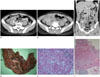
An abdominal computed tomography (CT) scan revealed a lobulated, peripherally enhancing, low density mass in the mesentery of the right lower quadrant, indicating necrotic lymphadenopathy (Fig. 1A). The patient underwent a laparoscopic biopsy to confirm the diagnosis. Laparoscopic excision was not feasible because of the presence of severe adhesions. Moreover, examination of intraoperative frozen sections revealed a granuloma, suggestive of tuberculosis. Histopathological examination showed chronic granulomatous inflammation with necrosis, and a polymerase chain reaction test for Mycobacterium tuberculosis was positive. The diagnosis was confirmed as tuberculous lymphadenitis, and anti-tuberculous treatment with isoniazid, rifampicin, ethambutol, and pyrazinamide was administered.
Symptoms improved initially, but after four months, the patient suddenly developed fever, chills, and lower abdominal pain. The patient underwent a follow-up CT scan for evaluation of the previous lymphadenopathy. The CT scan showed that the previous tuberculous lymphadenopathy had increased in size and now contained tiny air bubbles. Fistula formation was noted between the lymphadenopathy and the proximal ascending colon (Fig. 1B, C). The patient underwent laparoscopic right hemicolectomy. Histopathological examination revealed chronic granulomatous inflammation with necrosis associated with tuberculosis and its involvement with the intestinal wall, pericolic soft tissue, and lymph nodes (Fig. 1D-F). The fistulous tract between the tuberculous lymphadenopathy and the proximal ascending colon was also observed on the gross specimen. After a few weeks, the symptoms were relieved. They were attributed to the abscess and the fistula. The patient took anti-tuberculous medication for 6 months and was followed clinically.
DISCUSSION
In 2014, the number of newly notified patients with tuberculosis in the Republic of Korea was 34869 cases (68.7 patients per every 100000 persons) and about one-fifth of them (6963 cases; 13.7 patients per every 100000 persons) had extrapulmonary manifestations (35). Abdominal tuberculosis is the most common form of extrapulmonary tuberculosis, and lymphadenopathy is the most common manifestation of abdominal tuberculosis (4). On the CT scans, the majority with lymphadenitis showed enlarged lymph nodes with central low attenuation and a peripheral enhancing rim. Other observations include a conglomerate mass, enlarged homogeneous attenuated lymph nodes, and several small homogeneous lymph nodes. This case was considered primary intra-abdominal tuberculous lymphadenitis with no evidence of pulmonary tuberculosis.
Nagi et al. (6) reported that 7.6 percent of patients with gastrointestinal tuberculosis had localized perforation and fistula. Fistula may form anywhere near the site of gastrointestinal tuberculosis, but the most common location is the ileum. Rare cases of tuberculous fistulas have been seen in the Republic of Korea. A case of choledocho-duodenal fistula, two cases of intestinal tuberculosis with a duodenal fistula, and two cases of nododuodenal fistula have been reported (78).
In this case, the diagnosis was a confirmed tuberculous lymphadenitis by the initial biopsy examination, and clinical improvement with anti-tuberculous medication was expected. Although the symptoms were relieved initially, the patient subsequently developed severe abdominal pain. The follow-up CT scan showed the formation of an intra-abdominal abscess and fistula which were managed surgically.
Cho et al. (9) reported paradoxical responses in non-HIV-infected patients with peripheral lymph node tuberculosis. Although these patients did not have intra-abdominal tuberculous lymphadenitis, 20% of patients with lymphadenopathy in the cervical, submandibular, axillary and inguinal areas showed paradoxical responses after receiving anti-tuberculous medication. Increasing size of the enlarged lymph nodes, discharging sinuses, and new enlarged lymph nodes were noted. In this case, it was presumed that the tuberculous lymphadenitis, in the healing process, lead to the formation of the intra-abdominal abscess and the fistulous tract to the proximal ascending colon. This observation, corresponding to the two cases of nododuodenal fistula during anti-tuberculous treatment, was regarded as a paradoxical response of tuberculous lymphadenitis to the medication.
Generally, the mainstay of treatment for a fistula caused by intestinal tuberculosis is conservative management, including anti-tuberculous medication. Surgical management may be considered based on clinical symptoms. Spontaneous closure of the fistula may not occur even after four or six weeks of treatment (10).
In conclusion, this is a rare case of fistula formation, extending from intra-abdominal tuberculous lymphadenitis to the proximal ascending colon during anti-tuberculous treatment. To the best of our knowledge, there is no published literature describing a nodo-colonic fistula. It is crucial for radiologists to know the various manifestations of tuberculosis in a country where it is endemic. A tuberculous fistula could be cured by conservative treatment. Therefore, radiologists might advise surgeons regarding the treatment options, thus preventing unnecessary surgeries.
 XML Download