

 PDF
PDF ePub
ePub Citation
 Print
Print


INTRODUCTION
Fibromatosis is a benign tumor that is composed of fibrous tissue without apparent histologic features of malignancy. However, it has significant potential for local invasiveness and recurrence (1). Solitary intestinal fibromatosis is very rare: fewer than 7 cases of intraabdominal solitary fibromatosis arising from the colon have been reported in the English literature.
We report here a case of histologically confirmed intraabdominal solitary fibromatosis arising from the colon with an emphasis on computed tomography (CT) findings. To the best of our knowledge, this is the first report of such a case in Korea.
CASE REPORT
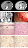
A 43-year-old female presented with a 2-day history of lower abdominal pain. The patient had undergone laparoscopic myomectomy 2 years ago. On physical examination, she was found to have diffuse abdominal tenderness, mainly in the epigastrium, with increased bowel sounds. Laboratory studies revealed a white blood cell count of 15.4 × 10³/uL and a C-reactive protein level of 1.2 mg/dL. A supine abdominal radiograph showed no gross abnormality. CT with contrast enhancement showed a well-circumscribed homogeneous ovoid-shaped mass in the splenic flexure of the colon, measuring 7 × 5 × 7 cm. The mass showed mild delayed enhancement [pre-contrast: 27 Hounsfield unit (HU), arterial phase: 40 HU, portal phase: 47 HU] (Fig. 1A, B). There was fat infiltration and edematous change in the greater omentum, mesentery, and splenic hilum, suggesting the likelihood of malignancy (Fig. 1C). Its imaging appearance was thought to be like that of a gastrointestinal stromal tumor. Esophagogastroduodenoscopy showed extrinsic compression along the greater curvature of the gastric body. Colonoscopy showed a 5 cm sized elevated lesion with normal mucosa at the splenic flexure of the colon, suggesting a submucosal tumor (Fig. 1D). Biopsy during colonoscopy only revealed colonic mucosa with lymphoid follicle but there was no evidence of malignancy.
On laparoscopic exam, there was a 7 cm sized submucosal mass arising from the splenic flexure of the colon with pericolic adhesion to the stomach, proximal jejunum, splenic hilum, and diaphragm. Laparoscopy-assisted left hemicolectomy, splenectomy, and stomach wedge resection were performed.
Histopathological evaluation of the excised colonic tumor showed a well-circumscribed, yellowish-white, firm and myxoid submucosal mass of the colon, measuring 7.5 × 7 × 4.8 cm in size. The tumor directly extended from the submucosa of the colon (Fig. 1E) to the splenic capsule and gastric proper muscle. The tumor was composed of spindle cell proliferation and finely collagenous stroma (Fig. 1F). Immunochemical stains showed immunopositivity for β-catenin (Fig. 1G) and smooth muscle actin (Fig. 1H), and negativity for the other markers (C-kit, CD34, S-100). The histopathological findings were consistent with deep fibromatosis.
DISCUSSION
Fibromatosis is a benign proliferation of fibrous tissue, characterized by infiltrative growth and frequent tendency to recur locally without metastatic potential. Fibromatoses are broadly categorized as superficial and deep on the basis of their anatomic location. Deep fibromatoses in the abdomen are classified as abdominal wall fibromatosis and intraabdominal fibromatosis (1). Intraabdominal solitary fibromatosis is extremely rare, and only 7 cases have occurred in the colon (Table 1). To the best of our knowledge, this is the first case report in Korea describing the CT findings of intraabdominal solitary fibromatosis arising from the colon. Familiarity with the imaging findings of fibromatosis is useful because complete resection may result in an apparent cure.
The majority of abdominal fibromatoses are sporadic. Among the 8 cases of solitary fibromatosis in the colon, this tumor was more commonly found in females (male:female ratio, 3:5) and the mean age at diagnosis showed a bimodal age distribution (40.5 days and 48.5 years) (2345678).
However, 9–18% of cases of mesenteric fibromatoses have a notable genetic association with familial adenomatous polyposis, specifically Gardner syndrome. Fibromatoses associated with Gardner syndrome are small and multiple and have a female predilection (ratio 3:1), whereas sporadic fibromatoses are generally larger and singular. Fibromatoses associated with Gardner syndrome tend to involve the mesentery and abdominal wall, whereas sporadic fibromatoses occur in the retroperitoneum and pelvis. In addition, Gardner syndrome-associated fibromatoses have a higher rate of recurrence after resection than do sporadic fibromatoses (9).
Imaging findings are variable and reflect the underlying histologic characteristics. On ultrasonogram, a lesion is visualized as a well-circumscribed or ill-defined solid mass with relatively homogenous hypoechogenicity with internal echogenic lines (10). CT is the best modality for establishing the diagnosis, determining the complications, and monitoring the treatment response. On CT, lesions with a highly collagenous stroma may be homogeneous and isoattenuating to muscle. Some lesions may appear striated or whorled when alternating collagenous and myxoid stroma is present. Contrast enhancement is variable; mild to moderate enhancement is typically noted, and delayed enhancement may be seen. Predominantly myxoid lesions do not enhance and remain hypoattenuating (234). On magnetic resonance (MR) imaging, most lesions show low to intermediate signal intensity on T1-weighted images; on T2-weighted images, the lesions can have heterogeneous intermediate or high signal intensity, and myxoid stromal elements contribute to high signal intensity. The enhancement pattern on MR imaging is variable, similar to the pattern observed on CT (10). The role of positron emission tomography-CT in evaluating fibromatoses remains undefined. 18F-fluorodeoxyglucose (FDG) uptake has been described in a segment of the colon affected by intussusception caused by solitary intestinal fibromatosis (maximum standardized uptake value = 5.4), although the inflammation associated with the intussusception precluded the assessment of FDG avidity of fibromatosis itself (3).
Differential diagnoses are gastrointestinal stromal tumor, inflammatory pseudotumor, and extrapleural solitary fibrous tumor. Other differential diagnoses include adenocarcinoma, leiomyosarcoma, plasmacytoma, and lymphoma.
The optimal therapeutic regimen for intraabdominal fibromatoses has not yet been determined. Some authors suggest that surgery is the first choice for treating fibromatoses and that the surgical aim is complete resection (10). Other authors recommend surgery only in cases that are complicated by bowel or ureteric obstruction. Some authors have suggested that chemotherapy is effective in aggressive cases of intraabdominal fibromatoses. When wide surgical margins cannot be achieved or when the tumor is not resectable, radiation therapy is generally recommended.
In conclusion, when confronted with a homogeneous submucosal mass arising from the colon that shows delayed mild enhancement, intraabdominal solitary fibromatosis should be included in the differential diagnosis.
 XML Download