

 PDF
PDF ePub
ePub Citation
 Print
Print


Abstract
Purpose
To compare the diagnostic utility of ultrasonography (US), CT and positron emission tomography/CT (PET/CT) in the preoperative evaluation of cervical lymph node metastasis in patients with papillary thyroid carcinoma.
Materials and Methods
The study population consisted of 300 patients with pathologically diagnosed papillary thyroid carcinoma after thyroidectomy and neck dissection. Preoperative US, CT, and PET/CT findings were compared with pathologic outcomes after thyroidectomy and neck dissection.
Results
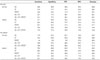
Sensitivity in detecting central lymph node metastasis (US 29.9%, CT 27.9%, PET/CT 18.8%) was lower than that for lateral lymph node metastasis (US 56.3%, CT 66.2%, PET/CT 43.7%). Specificity in detecting central lymph node metastasis (US 80.6%, CT 77.7%, PET/CT 83.0%) was lower than that for lateral lymph node metastasis (US 96.8%, CT 80.6%, PET/CT 95.2%). The combination of US and CT had higher specificity (77.3%) and higher sensitivity (33.1%) than US alone. PET/CT has no significant additional benefit over the combination of US and CT.
Figures and Tables
Fig. 1
US image of a 64-year-old female with papillary thyroid carcinoma. A hyperechoic lymph node is shown (between +) in the right internal jugular chain. Histopathological analysis revealed a metastatic papillary carcinoma.
US = ultrasonography
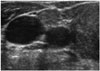
Fig. 2
US image of a 49-year-old male with papillary thyroid carcinoma. Microcalcification (between +) is shown (+) in the left lateral neck lymph node. Histopathological analysis revealed metastatic papillary carcinoma.
US = ultrasonography
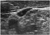
Fig. 3
US image of a 66-year-old male with papillary thyroid carcinoma. Cystic changes (between +) are observed in the lymph node. Histopathological analysis revealed metastatic papillary carcinoma.
US = ultrasonography
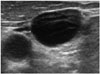
Fig. 4
CT image of a 62-year-old male with papillary thyroid carcinoma. The right paratracheal lymph node is increased in size and shows calcification (arrow). Histopathological analysis revealed metastatic papillary carcinoma.

Table 1
Patient Demographics and Tumor Characteristics (n= 300)
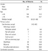
Table 2
Numbers of Dissected Nodes and Positive Neck Nodes per Level and per Patient
Table 3
Sensitivity, Specificity, PPV, NPV, and Diagnostic Accuracy of Preoperative US, CT, and PET/CT per Level and per Patient (%)
References
1. Shaha AR, Shah JP, Loree TR. Patterns of nodal and distant metastasis based on histologic varieties in differentiated carcinoma of the thyroid. Am J Surg. 1996; 172:692–694.
2. Hundahl SA, Fleming ID, Fremgen AM, Menck HR. A national cancer data base report on 53,856 cases of thyroid carcinoma treated in the U.S., 1985-1995 [see commetns]. Cancer. 1998; 83:2638–2648.
3. DeGroot LJ. Long-term impact of initial and surgical therapy on papillary and follicular thyroid cancer. Am J Med. 1994; 97:499–500.
4. McConahey WM, Hay ID, Woolner LB, van Heerden JA, Taylor WF. Papillary thyroid cancer treated at the Mayo Clinic, 1946 through 1970: initial manifestations, pathologic findings, therapy, and outcome. Mayo Clin Proc. 1986; 61:978–996.
5. Coburn MC, Wanebo HJ. Prognostic factors and management considerations in patients with cervical metastases of thyroid cancer. Am J Surg. 1992; 164:671–676.
6. Hughes CJ, Shaha AR, Shah JP, Loree TR. Impact of lymph node metastasis in differentiated carcinoma of the thyroid: a matched-pair analysis. Head Neck. 1996; 18:127–132.
7. Lee KD, Lee HS. Central neck dissection for papillary thyroid carcinoma. J Korean Thyroid Assoc. 2014; 7:140–148.
8. Rosário PW, de Faria S, Bicalho L, Alves MF, Borges MA, Purisch S, et al. Ultrasonographic differentiation between metastatic and benign lymph nodes in patients with papillary thyroid carcinoma. J Ultrasound Med. 2005; 24:1385–1389.
9. Ying M, Ahuja A, Metreweli C. Diagnostic accuracy of sonographic criteria for evaluation of cervical lymphadenopathy. J Ultrasound Med. 1998; 17:437–445.
10. Na DG, Lim HK, Byun HS, Kim HD, Ko YH, Baek JH. Differential diagnosis of cervical lymphadenopathy: usefulness of color Doppler sonography. AJR Am J Roentgenol. 1997; 168:1311–1316.
11. Som PM, Brandwein M, Lidov M, Lawson W, Biller HF. The varied presentations of papillary thyroid carcinoma cervical nodal disease: CT and MR findings. AJNR Am J Neuroradiol. 1994; 15:1123–1128.
12. Mitchell JC, Grant F, Evenson AR, Parker JA, Hasselgren PO, Parangi S. Preoperative evaluation of thyroid nodules with 18FDG-PET/CT. Surgery. 2005; 138:1166–1174. discussion 1174-1175.
13. Som PM. Detection of metastasis in cervical lymph nodes: CT and MR criteria and differential diagnosis. AJR Am J Roentgenol. 1992; 158:961–969.
14. Stulak JM, Grant CS, Farley DR, Thompson GB, van Heerden JA, Hay ID, et al. Value of preoperative ultrasonography in the surgical management of initial and reoperative papillary thyroid cancer. Arch Surg. 2006; 141:489–494. discussion 494-496.
15. Shimamoto K, Satake H, Sawaki A, Ishigaki T, Funahashi H, Imai T. Preoperative staging of thyroid papillary carcinoma with ultrasonography. Eur J Radiol. 1998; 29:4–10.
16. Jeong HS, Baek CH, Son YI, Choi JY, Kim HJ, Ko YH, et al. Integrated 18F-FDG PET/CT for the initial evaluation of cervical node level of patients with papillary thyroid carcinoma: comparison with ultrasound and contrast-enhanced CT. Clin Endocrinol (Oxf). 2006; 65:402–407.
17. Ito Y, Tomoda C, Uruno T, Takamura Y, Miya A, Kobayashi K, et al. Clinical significance of metastasis to the central compartment from papillary microcarcinoma of the thyroid. World J Surg. 2006; 30:91–99.
18. Ito Y, Uruno T, Nakano K, Takamura Y, Miya A, Kobayashi K, et al. An observation trial without surgical treatment in patients with papillary microcarcinoma of the thyroid. Thyroid. 2003; 13:381–387.
19. Ahuja A, Ying M. Sonography of neck lymph nodes. Part II: abnormal lymph nodes. Clin Radiol. 2003; 58:359–366.
20. Kuna SK, Bracic I, Tesic V, Kuna K, Herceg GH, Dodig D. Ultrasonographic differentiation of benign from malignant neck lymphadenopathy in thyroid cancer. J Ultrasound Med. 2006; 25:1531–1537. quiz 1538-1540.
21. van den Brekel MW, Castelijns JA, Snow GB. The size of lymph nodes in the neck on sonograms as a radiologic criterion for metastasis: how reliable is it? AJNR Am J Neuroradiol. 1998; 19:695–700.
22. Son KR, Na DG, Chang KH. Diagnostic value of CT for the detection of cervical lymph node metastases in papillary thyroid carcinoma. J Korean Soc Radiol. 2009; 60:383–389.
23. Verge J, Guixá J, Alejo M, Basas C, Quer X, De Castro J, et al. Cervical cystic lymph node metastasis as first manifestation of occult papillary thyroid carcinoma: report of seven cases. Head Neck. 1999; 21:370–374.
24. Hwang CF, Wu CM, Su CY, Cheng L. A long-standing cystic lymph node metastasis from occult thyroid carcinoma--report of a case. J Laryngol Otol. 1992; 106:932–934.
25. Cooper DS, Doherty GM, Haugen BR, Kloos RT, Lee SL, Mandel SJ, et al. Management guidelines for patients with thyroid nodules and differentiated thyroid cancer. Thyroid. 2006; 16:109–142.
26. Amdur RJ, Mazzaferri EL. Essentials of Thyroid Cancer Management. New York: Springer;2005. p. 211–214.
27. Costa A, Testori OB, Cenderelli C, Giribone G, Migliardi M. Iodine content of human tissues after administration of iodine containing drugs or contrast media. J Endocrinol Invest. 1978; 1:221–225.
28. Spate VL, Morris JS, Nichols TA, Baskett CK, Mason MM, Horsman TL, et al. Longitudinal study of iodine in toenails following IV administration of an iodine-containing contrast agent. J Radioanal Nucl Chem. 1998; 236:71–77.
29. Kim E, Park JS, Son KR, Kim JH, Jeon SJ, Na DG. Preoperative diagnosis of cervical metastatic lymph nodes in papillary thyroid carcinoma: comparison of ultrasound, computed tomography, and combined ultrasound with computed tomography. Thyroid. 2008; 18:411–418.
30. Lee DW, Ji YB, Sung ES, Park JS, Lee YJ, Park DW, et al. Roles of ultrasonography and computed tomography in the surgical management of cervical lymph node metastases in papillary thyroid carcinoma. Eur J Surg Oncol. 2013; 39:191–196.
31. Joo YH, Yun CH, Cho JH, Sun DI, Yoo IR, Jung SR, et al. Utility of FDG PET/CT scans on nodal staging in patients with papillary thyroid cancer. J Korean Thyroid Assoc. 2008; 1:107–111.
 XML Download