

 PDF
PDF ePub
ePub Citation
 Print
Print


Abstract
Materials and Methods
691 patients visited the emergency department of our hospital between January 2006 and October 2012 for paraquat poisoning. Of these 691, we identified 56 patients whose initial chest radiographs were normal but who developed radiographic abnormalities within one week. We evaluated their radiographic findings and the differences in imaging features based on mortality.
Results
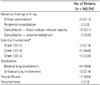
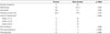
The most common finding was diffuse consolidation (29/56, 52%), followed by consolidation with linear and nodular opacities (18/56, 32%), and combined consolidation and pneumomediastinum (7/56, 13%). Pleural effusion was noted in 17 patients (30%). The two survivors (4%) showed peripheral consolidations, while the 54 patients (96%) who died demonstrated bilateral (42/54, 78%) or unilateral (12/54, 22%) diffuse consolidations.
Figures and Tables
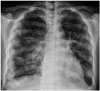 | Fig. 1A 33-year-old female who ingested 50 mL of paraquat. Chest radiograph obtained 4 days after ingestion reveals rapidly progressing diffuse consolidation in both lungs.
|
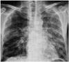 | Fig. 2A 55-year-old male who ingested 30 mL of paraquat. Chest radiograph performed 5 days after ingestion shows diffuse consolidation in both lungs with reticular density in the right lower lung.
|
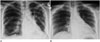 | Fig. 3A 45-year-old female who survived after ingesting 15 mL of paraquat.
A. 6-day follow-up radiograph reveals peripheral consolidation in both lower lungs.
B. 6-month follow-up radiograph demonstrates resolution of previous peripheral consolidation in both lower lungs, with minimal residual fibrosis.
|
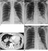 | Fig. 4A 47-year-old female who survived after ingesting 30 mL of paraquat.
A. 3-day follow-up radiograph shows subtle opacities in both basal lungs.
B. 8-day follow-up image demonstrates aggravated multifocal consolidations in both lungs, especially the perepheral portions.
C. Chest CT scan obtained 7 weeks after ingestion shows consolidation and dilatation of bronchial structures from fibrotic change.
D. One year follow-up image shows residual fibrosis in both lungs.
|
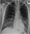 | Fig. 5A 33-year-old female who ingested 350 mL of paraquat. Chest radiograph obtained 3 days after ingestion shows diffuse consolidation in bilateral middle and lower lungs with pneumomediastinum.
|
Table 1
Summary of Chest Radiographic Findings of Paraquat Poisoning
![]()
Table 2
Frequency and Distribution of Abnormality on Chest Radiographs in 56 Patients with Paraquat Poisoning
![]()
References
1. Davies DS, Hawksworth GM, Bennett PN. Paraquat poisoning. Proc Eur Soc Toxicol. 1977; 18:21–26.
2. Gawarammana IB, Buckley NA. Medical management of paraquat ingestion. Br J Clin Pharmacol. 2011; 72:745–757.
3. Lee KH, Gil HW, Kim YT, Yang JO, Lee EY, Hong SY. Marked recovery from paraquat-induced lung injury during long-term follow-up. Korean J Intern Med. 2009; 24:95–100.
4. Im JG, Lee KS, Han MC, Kim SJ, Kim IO. Paraquat poisoning: findings on chest radiography and CT in 42 patients. AJR Am J Roentgenol. 1991; 157:697–701.
5. Bullivant CM. Accidental poisoning by paraquat: report of two cases in man. Br Med J. 1966; 1:1272–1273.
6. Matthew H, Logan A, Woodruff MF, Heard B. Paraquat poisoning--lung transplantation. Br Med J. 1968; 3:759–763.
7. Suntres ZE. Role of antioxidants in paraquat toxicity. Toxicology. 2002; 180:65–77.
8. Rose MS, Lock EA, Smith LL, Wyatt I. Paraquat accumulation: tissue and species specificity. Biochem Pharmacol. 1976; 25:419–423.
9. Sabzghabaee AM, Eizadi-Mood N, Montazeri K, Yaraghi A, Golabi M. Fatality in paraquat poisoning. Singapore Med J. 2010; 51:496–500.
10. Lee SH, Lee KS, Ahn JM, Kim SH, Hong SY. Paraquat poisoning of the lung: thin-section CT findings. Radiology. 1995; 195:271–274.
11. Kim YT, Kim HC, Bae WK, Kim IY, Im HH. Paraquat induced lung injury: long-term follow-up of HRCT. J Korean Radiol Soc. 2004; 50:179–183.
 XML Download