

 PDF
PDF ePub
ePub Citation
 Print
Print


INTRODUCTION
Leiomyomas are common benign tumors. They are present in about 20% of women of reproductive age (1). Large uterine fibroids are associated with infertility, menorrhagia, pain, and mass effects (2). The most common site of leiomyoma is the uterus, but they can be found in the broad ligaments, ovaries, and vagina (23). Abdominal wall leiomyomas are rare, and likely result from seeding following the surgical resection of uterine leiomyomas, especially laparoscopic surgery (34). We presented a very rare case of solitary leiomyoma of the anterior abdominal wall in a patient without antecedent gynecological surgeries. We described the imaging findings of this tumor because they are rarely reported. This report was approved by our Institutional Review Board.
CASE REPORT
A 33-year-old female with gravida 2 para 2 presented with a palpable abdominal mass in the left lower quadrant. The patient did not complain of any symptoms or signs except the palpable mass. There was no history of steroid usage, hormone replacement therapy, previous surgery or gynecologic interventions. A smooth, non-tender, movable pre-peritoneal mass that measured about 1 to 2 cm was detected on physical examination. It had a smooth surface and was not attached to the overlying skin.
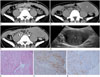
The computed tomography (CT) of the abdomen revealed thickening of the left rectus abdominis muscle at the lower level of the umbilicus and the loss of intervening fat between the rectus abdominis muscle and the lateral abdominal wall muscles (Fig. 1A). It was similarly enhanced in the adjacent muscles. The uterus and both ovaries showed no pathologic findings.
After 8 months, the patient complained that the size of the left abdominal wall mass had increased. A follow-up CT scan and ultrasound were performed to evaluate the mass. The CT scan showed a lentiform-shaped mass measuring 10.4 × 5.3 × 6.1 cm in the left rectus abdominis muscle involving the left lateral abdominal muscles. On the non-contrast image, the mass had a well-circumscribed margin and hypodensity, as compared with the adjacent muscles (Fig. 1B). It was homogeneously enhanced and showed isodensity near the adjacent muscles (Fig. 1C). The left lateral abdominal wall muscles were displaced laterally by the mass effects of the mass. There was no invasion of the peritoneal cavity or intra-abdominal visceral organs.
The ultrasound showed a relatively well-defined and mixed hypoechoic mass with a lentiform shape in the left rectus abdominis muscle (Fig. 1D). The patient subsequently underwent ultrasonography-guided needle biopsy at multiple sites. Histopathology showed a probably benign spindle cell tumor without mitosis, necrosis, or atypia. The spindle cells were positive for smooth muscle actin, partly for desmin, and negative for CD 68 and S100. These features were consistent with a benign leiomyoma (Fig. 1E-G).
DISCUSSION
Leiomyoma of the anterior abdominal wall is rare. It is thought to usually occur with the seeding of myomatous tissue during previous gynecological surgeries, and is more common in laparoscopic procedures than in laparotomy (5). Thus, there are few reported cases of isolated abdominal wall leiomyoma without previous gynecologic surgeries or the presence of uterine fibroids. The diagnosis of de novo leiomyoma of the abdominal wall can be made even though the patient has no history of previous abdominal surgery, as was the case in our patient. Theoretically, a leiomyoma can be found anywhere where there are smooth muscles, including the abdominal wall. De novo leiomyoma can even develop in areas devoid of myomatous tissues or obvious smooth muscle cells (6). Therefore, leiomyoma should be included in the list of the differential diagnoses for abdominal wall mass.
Abdominal wall leiomyomas can show the loss of intervening fat between the rectus abdominis and lateral abdominal wall muscles, and they are usually hypodense without significant contrast enhancement (78). Our case was also hypodense to the adjacent muscles and intermediately enhanced. However, because abdominal wall endometriosis and desmoid tumors are relatively more enhanced than leiomyomas, they are usually hyperdense, as compared with the muscles (910). A helpful defining feature is that leiomyomas are usually lentiform-shaped masses, whereas endometriosis and desmoid tumors are usually round or oval masses. Sarcomas, such as rhabdomyosarcomas, fibrosarcomas, leiomyosarcomas and hematomas, could present as abdominal wall masses. Sarcomas tend to be heterogeneously enhanced, show necrotic areas, and invade adjacent structures. In our case, the biopsy was performed at multiple sites; and the imaging findings were suggestive of a low possibility of malignancy. Patients with hematoma usually have a typical history, such as trauma, surgery, or anticoagulation. In addition, the typical location and configuration or hematocrit effect could be helpful in a differential diagnosis.
In conclusion, abdominal wall leiomyoma is a rare presentation. It usually occurs after a previous abdominal surgery, but can develop de novo in a fertile woman. Therefore, a leiomyoma should be included in the differential diagnosis of the abdominal wall mass even in a patient without previous surgery. In addition, we suggest some radiologic findings that could be helpful in the differential diagnosis of abdominal wall masses prior to histology.
 XML Download