

 PDF
PDF ePub
ePub Citation
 Print
Print


INTRODUCTION
Retroperitoneal fibrosis (RPF) is an uncommon fibrotic reaction in the retroperitoneum. There is an idiopathic form and a secondary form associated with IgG4-related disease, drugs (methysergide, pergolide, bromocriptine, ergotamine, methyldopa, hydralazine), infections (tuberculosis, histoplasmosis, actinomycosis), malignancies (carcinoid tumor, Hodgkin's and non-Hodgkin's lymphomas, sarcomas, carcinomas of the colon, prostate, breast, and stomach), prior surgeries, and radiation therapy. The idiopathic form accounts for more than two-thirds of cases and it was thought to result from a local inflammatory response to antigens in the atherosclerotic plaques in the abdominal aorta, combined with auto-immunologic factors. Thus, the fibroinflammatory tissue usually encases the infrarenal portion of the abdominal aorta, inferior vena cava, and ureters (123). Here, we report two rare cases of idiopathic RPF involving only a unilateral renal pelvis and sinus; thus mimicking renal pelvic cancer.
CASE REPORT
Case 1
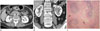
A 66-year-old female presented with an incidental finding at computed tomography (CT) screening. Her previous medical history revealed that she had undergone hysterectomy for an unknown cause 10 years ago. Contrast-enhanced genitourinary CT showed a homogeneously enhancing soft tissue mass replacing the left renal pelvis, which was suspicious for renal pelvic cancer. The renal pelvis was slightly dilated, but ureteric dilatation was not present (Fig. 1A, B).
Urine cytologies of voided urine and washed urine from the left renal pelvis were negative.
On retrograde pyelography (RGP) performed by urologists, there was no mass-like lesion and biopsy of the left renal pelvis also yielded unsatisfactory results. She underwent laparoscopic radical nephrectomy. Histopathologic findings of the resected kidney were negative for malignancy. Fibrosis and chronic inflammation with myofibroblast proliferation and lymphoplasma cell infiltration were observed. The lesion was mainly located in the renal sinus fat tissue around renal vessels, suggesting an inflammatory or immunologic process such as idiopathic RPF (Fig. 1C). Immunochemistry of IgG and IgG4 was performed to confirm the presence of IgG4+ plasma cells in the tissue. The ratio of IgG4 to IgG plasma cells was within the normal range (< 5%), and these results were inconsistent with the diagnosis of IgG4-related disease, which shows an IgG4 to IgG ratio of more than 30-40%. Pathology report was compatible with idiopathic RPF.
Case 2
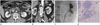
An 80-year-old man presented with fever and cough for one week. His past history was nonspecific except for hypertension. There was no abnormal finding on physical examination. The results of peripheral blood smear examination showed slightly elevated C-reactive protein (2.93 mg/dL). On chest CT, mild bronchitis and the incidental finding of a left renal pelvic mass were detected. Additional contrast enhanced genitourinary CT was performed for further evaluation. CT showed an enhancing soft tissue mass in the left renal pelvis and sinus with mild pelvic dilatation. Periureteric fat infiltration in the left proximal ureter without definite wall thickening was observed (Fig. 2A, B).
Two urine cytologies of washed urine from the left renal pelvis were negative, and on RGP, there was no filling defect but mild luminal narrowing of the renal pelvis and mild calyectasia were detected (Fig. 2C). He underwent laparoscopic radical nephrectomy. Histopathologic findings were consistent with idiopathic RPF (Fig. 2D). Stromal fibrosis with lymphoplasma cell infiltration mainly into the renal sinus was observed. On immunochemistry of the tissue, the ratio of IgG4 to IgG plasma cells was less than 5%, which was within the normal range, and on laboratory examination, serum levels of IgG and IgG4 were within their normal range [1056.6 mg/dL (range, 700-1600 mg/dL) and 1020.0 mg/L (range, 30-2010 mg/L), respectively].
DISCUSSION
RPF is a rare autoimmune, inflammatory disorder that develops in about one in 200000 people (4). It is most common in individuals 40 to 60 years of age (5).
Men are affected by this disorder two to three times more often than women (1). In typical RPF, a mass of fibrous tissue develops around the abdominal aorta with an epicenter at the L4-5 level and it also encases the inferior vena cava and the ureter. Ureteral involvement is reported in 80-100% of cases at presentation (4). At presentation, such ureteral involvement is often bilateral, but in patients with an apparently unilateral obstruction, contralateral disease can develop even within a short period (1).
CT is the modality of choice for imaging diagnosis of RPF and for evaluating the extent of the process. On CT scan, RPF usually appears as a homogeneous plaque, which is isodense to the adjacent muscle, surrounding the lower abdominal aorta and the iliac arteries, and often enveloping the ureters and the inferior vena cava (4). The degree of contrast enhancement is related to inflammation and process activity.
Intense enhancement indicates fibrous tissue in an initial phase with associated inflammation (6). The advantage of MR imaging is avoidance of nephrotoxicity of iodinated contrast media in patients with compromised renal function and better definition against the surrounding tissues, mainly when fat-saturation images are used.
Idiopathic RPF is hypointense in T1-weighted MR images. In T2-weighted MR images, its intensity is variable; high signal intensity in the active inflammatory stage because of tissue edema and hypercellularity and low signal intensity in the late fibrotic stage (567).
In rare cases, RPF presents as an asymmetric poorly circumscribed retroperitoneal mass with atypical localization, and thus it is not characterized by aortic involvement as observed in our cases, and differentiation from a primary retroperitoneal tumor or malignant lymphadenopathy is very difficult. Atypical sites of RPF involvement include the small bowel mesentery, duodenum, colon, bladder, and periduodenal, peripancreatic, pelvic, periureteral and epidural space (14).
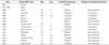
Perirenal extension of RPF is rarely reported (678910), whereas in only a few cases, the disease was unilaterally localized. To the best of our knowledge, three cases of idiopathic RPF presenting with features of a localized unilateral renal pelvic mass mimicking renal pelvic cancer (89) without periaortic or pericaval involvement have been reported (Table 1).
IgG4-related kidney disease is an important part of the spectrum of IgG4-related sclerosing disease, which manifests as a secondary form of RPF. IgG4-related kidney disease is usually accompanied by other organ involvement, and isolated IgG4-related kidney disease without other organ involvement is very rare; only 6% of cases have been reported as renal lesions alone (10).
Although CT and MR imaging have an important role in diagnosis of RPF, histological tissue examination is needed, especially in cases with atypical location of a retroperitoneal mass to rule out malignancy. In addition to malignancy (urothelial carcinoma), differential diagnosis includes retroperitoneal fibromatosis, which is characterized by uniform proliferation of fibroblasts and is associated with Gardner's syndrome. Another differential diagnosis is inflammatory pseudotumor, which mainly affects children, and its appearance is that of a huge mass with infiltrative borders. It is histologically characterized by myofibroblast proliferation with myxoid and inflammatory areas (1).
 XML Download