

 PDF
PDF ePub
ePub Citation
 Print
Print


INTRODUCTION
Fibrosing mediastinitis is a rare benign disorder that is characterized by deposition of dense fibrous tissue throughout the mediastinum and hilar regions (1). Resultant infiltrative or compressive effects on vital mediastinal organs such as central systemic vein, airway, and pulmonary vessels, could increase morbidity and in rare circumstances even mortality (2). We reported a rare case of fibrosing mediastinitis causing pulmonary infarction by encasing pulmonary artery that was detected on chest computed tomography (CT).
CASE REPORT
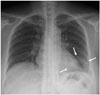
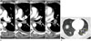
A 44-year-old female patient visited the emergency room (ER) of a tertiary referral university hospital due to acute hemoptysis and right chest wall pain of 2 months duration. Her chest pain was refractory to prior conservative treatment in a local clinic. The patient was a nonsmoker and had no particular medical history. On laboratory tests, serum erythrocyte sedimentation rate was mildly elevated but other parameters were within normal limits. Plain chest radiograph demonstrated patchy increased opacity in left lower lung zone and small amount of left pleural effusion (Fig. 1). Considering the refractory nature of her chest pain and the acute onset of hemoptysis, post-contrast chest CT was immediately conducted in the ER. Chest CT revealed soft tissue mass at the left hilar region, which showed homogenous enhancement and was encasing and provoking near total occlusion of left inferior pulmonary artery (Fig. 2A). There was no visible calcification within the mass. Wedgeshaped subpleural consolidation was seen in the left lower lobe, which was the corresponding vascular territory of the encased left lower pulmonary artery (Fig. 2B). Therefore, this consolidative lesion could be regarded as pulmonary infarction. Laboratory screenings with antinuclear antibody, rheumatoid factor, and Anti-neutrophil cytoplasmic antibody were negative.
Without further investigation such as positron emission tomography-CT or bronchoscopy, open thoracotomy biopsy was performed for confirmative diagnosis owing to the patient's severe symptoms. Extensive left peribronchial fibrosis and soft tissue mass encasing left inferior pulmonary artery was seen intra-operatively. Except for superior segment, the remaining entire left lower lobe was rigid on palpation and there were multifocal scattered infarctions.
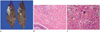
Gross examination of resected specimen revealed firm white fibrous tissue surrounding the left hilum (Fig. 3A). Histopatho-logically, the mass consisted of dense fibrosis surrounding and infiltrating pulmonary vasculature with extension into the lung parenchyme (Fig. 3B). Occasional chronic necrotizing granulomas were seen within the dense fibrosis (Fig. 3C). Polymerase chain reaction (PCR) for tuberculosis was negative and Gomori methenamine silver (GMS) stain for fungal study was also negative. Consequently, the final diagnosis was fibrosing mediastinitis. The patient underwent follow-up for 3 years and was free from symptom and recurrence on imaging.
DISCUSSION
Fibrosing mediastinitis is a benign progressive non-neoplastic disease characterized by infiltration of excessive fibrous tissue within the mediastinum mediated by abnormal immune reaction to various antigens or underlying disease (3). Fibrosing mediastinitis is interchangeably known as mediastinal granuloma and mediastinal fibrosis, sclerosing mediastinitis, and granulomatous mediastinitis, based on dominant pathologic features (4). Although it is still controversial, Loyd et al. (5) postulated that granulomatous mediastinitis evolves into fibrosing mediastinitis; hence, granulomatous mediastinitis and fibrosing mediastinitis may represent 2 ends of a spectrum of abnormal inflammation and fibroproliferation in the mediastinum (6).
In our case, pathologic examination indicated extensive dense fibrosis surrounding and infiltrating pulmonary vasculature with extension into the lung parenchyme and occasional chronic necrotizing granulomas within the dense fibrosis. Thus, our case showed both dominant features of fibrosing mediastinitis and minor features of granulomatous mediastinitis.
Fibrosing mediastinitis is most commonly associated with histoplasma infection in North America (5). Other possible triggers of fibrosing mediastinitis are reportedly tuberculosis, fungal infections, and sarcoidosis (6). However, if certain identifiable cause is absent, the disease is categorized as idiopathic (6). In many cases, no specific microorganism is identified and thus categorized as idiopathic (7). In our case, laboratory examination and elastin stain for vasculitis were negative and investigation for immunoglobulin G4-related disease was also negative. In addition, PCR test for tuberculosis and non-tuberculosis mycobacterium were negative, as well as GMS tests for fungal study. Therefore, our case could be categorized as idiopathic fibrosing mediastinitis.
Fibrosing mediastinitis may present with various clinical manifestations according to the anatomical location involved. While some authors have reported that bronchial narrowing and superior vena cava (SVC) obstruction are the most common complication of fibrosing mediastinitis (8), others have reported pulmonary artery stenosis followed by SVC narrowing as the most common and significant manifestation (3). On CT scan, fibrosing mediastinitis is characterized by localized or diffuse softtissue mass in mediastinum obliterating fat planes and causing compression of adjacent structures. Additional abnormal findings associated with disease localization within the pleura and lungs could include pulmonary infarction from pulmonary artery obstruction, as in our case (3). CT can accurately identify tracheobronchial narrowing, major vessel obstruction, and degree of pulmonary arterial hypertension. In our patient, CT demonstrated the typical findings of fibrosing mediastinitis. Differential diagnosis included other mediastinal mass forming diseases such as Castleman's disease and lymphoma. The mass was non-calcified soft tissue mass and surgical tissue acquisition was necessary to exclude neoplastic diseases.
The treatment of fibrosing mediastinitis is controversial. The effect of systemic antifungal or antituberculous therapy is reported in cases with confirmed causative microorganism (9). However, in cases of granuloma or fibrosis causing compression of mediastinal organ, surgical resection of affected tissue is necessary (10). In our case, the patient suffered from chest wall pain and blood tinged sputum as a result of pulmonary infarction. Surgical resection was necessary for symptom relief as well as histopathologic diagnosis. Consequently, the patient's symptoms were completely resolved for the following 3 years follow up period.
Our case demonstrated the rare occurrence of fibrosing mediastinitis with pulmonary artery occlusion and implication of surgical resection for local disease control.
 XML Download