

 PDF
PDF ePub
ePub Citation
 Print
Print


INTRODUCTION
Ultrasonography is an accurate method for the detection of thyroid nodules, but it has limited utility in differentiating between benign and malignant thyroid nodules (1). Fine-needle aspiration (FNA) is a standard diagnostic procedure performed to obtain tissue for pathologic examination to determine whether a thyroid nodule is malignant. FNA is known to have a high specificity (60–98%) but varying sensitivity (54–90%) for the diagnosis of malignant thyroid nodules (2345). A classical criterion of malignancy is a nodule that is hard or firm upon palpation or with ultrasonography probe pressure (6). Therefore, assessment of tissue stiffness has become a viable method to differentiate thyroid nodules. Acoustic radiation force impulse (ARFI) imaging is an ultrasonographic elasticity imaging technology that noninvasively assesses tissue elasticity and quantifies the measured tissue stiffness as a velocity of ARFI. The quantitative application of ARFI technology is called virtual touch tissue quantification (VTQ) technology. The stiffer a tissue is, the greater the VTQ velocity is (7). VTQ has been applied to help distinguish between benign and malignant lesions in the breast, liver, and kidney (8910111213). More recently, studies have also been performed to investigate the application of VTQ technology to the thyroid (14151617); however, VTQ needs further testing before it can be utilized as a clinical imaging tool.
The aim of the present study was to evaluate the diagnostic utility of VTQ technology in differentiating between benign and malignant thyroid nodules.
MATERIALS AND METHODS
Patients
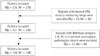
This retrospective study was approved by the Institutional Review Board of our hospital, and the requirement for informed consent was waived. The study period was from September 2012 to August 2013. A total of 198 thyroid nodules in 164 consecutive patients (24 men and 140 women; mean age 53.8 ± 12.1 years, range 18–76 years) were retrospectively examined by one radiologist with 10 years of experience in thyroid ultrasonography and FNA. The flow chart for patient selection is shown in Fig. 1.
The final inclusion criteria were as follows: 1) nodules with a solid component that was 0.5 cm or greater in diameter; 2) sufficient adjacent thyroid tissue surrounding the nodule at the same depth in the region of interest (ROI); 3) nodules with an available VTQ velocity (0–10 m/s); and 4) nodules with a benign or malignant FNA cytology or post-surgical pathological results.
Nodules with Bethesda category I, III, IV, or V without postsurgical pathological results were excluded.
Conventional Ultrasonography
Conventional ultrasonography was done with an Acuson S2000 ultrasound system (Siemens Medical Solutions, Mountain View, CA, USA) equipped with a linear array transducer (bandwidth frequency of 4–9 MHz) and ARFI software. Patients were positioned in the supine position with a fully exposed neck. The morphologic characteristics, size, boundary, and echogenicity of the nodule were observed by conventional ultrasonography.
Acoustic Radiation Force Impulse Imaging
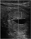
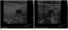
Switching to the ARFI mode, VTQ was utilized to measure the stiffness of the nodules and the adjacent normal thyroid tissues. The ROI measuring 0.5 × 0.6 cm was placed within the solid portion of the nodules. When a solid-and-cystic nodule was encountered, the ROI was centered within the solid component, and the FNA was also performed at the same location (Fig. 2). In cases of a large nodule with a heterogeneous solid portion, the ROI was placed in the most hypoechoic portion of the nodule. The VTQ velocity measurement was taken five times and completed within 3 minutes in most cases. The results were expressed in m/s with a measurement range of 0–10 m/s. The median value was selected as the VTQ velocity of the nodule. VTQ velocities greater than 10 m/s were not able to be evaluated by this ultrasound system and were displayed as 'x.xx m/s'. In cases without five valid measurements (numeric values), and more 'x.xx m/s' measurements than numeric values, the nodule was repeatedly measured to get an adequate result. VTQ measurements of adjacent normal thyroid tissue were also taken at the same depth and in the same manner as that of the nodules. In cases of heterogeneous thyroid parenchyma, such as thyroiditis, the VTQ velocity was measured in the same way as normal parenchymal tissue measurements. The VTQ velocity ratio between each nodule and the adjacent thyroid tissue was also calculated.
FNA and Histopathology
FNA was performed by the same radiologist who examined the conventional ultrasonography and ARFI imaging with a 21- or 25-gauge needle. The FNA result was reported according to the Bethesda system for reporting thyroid cytopathology (18). For a thyroid FNA specimen to be satisfactory for evaluation, at least 6 groups of benign follicular cells were required with each group composed of at least 10 cells. Non-diagnostic results were applied to specimens that were unsatisfactory due to obscuring blood, overly thick smears, air drying of alcohol-fixed smears, or an inadequate number of follicular cells. FNA cytology or post-surgical histopathological results were used as the standard of reference for the diagnosis of a benign or malignant thyroid nodule.
Statistical Analysis
All measured data were expressed as the mean value ± standard deviation. A p value of less than 0.05 was considered as statistically significant. The VTQ velocities and VTQ velocity ratio between benign and malignant groups were compared by Student's t-test with SPSS 20 (SPSS Inc., Chicago, IL, USA). Receiver operating characteristic curve was used to assess the diagnostic performance of the VTQ velocity and VTQ velocity ratio. As a result, the best cutoff value was determined with MedCalc ver. 13.1.2.0 (MedCalc software, Mariakerke, Belgium).
RESULTS
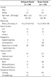
In total, 168 benign nodules and 30 malignant nodules were included in this study. Demographic features of the patients and ultrasonographic features of the thyroid nodules are presented in Table 1. In 168 benign nodules, there were 166 benign follicular lesions and 2 cases of thyroiditis (1 Hashimoto thyroiditis and 1 lymphocytic thyroiditis). All 30 malignant nodules were papillary thyroid carcinoma.
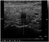
Malignant nodules had significantly higher VTQ velocities (3.06 ± 1.04 m/s, range: 1.90–6.46 m/s) (Fig. 3) than those of benign nodules (2.40 ± 0.85 m/s, range: 0.69–8.09 m/s) (p = 0.002). The optimal cutoff velocity was 2.37 m/s for accurate differentiation between benign and malignant thyroid nodules. The area under the receiver operating characteristic curve was 0.72. The sensitivity, specificity, positive predictive value, negative predictive value, and accuracy were 86.7%, 50.6%, 23.9%, 95.5%, and 56.1%, respectively.
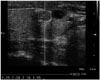
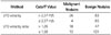
The VTQ velocity ratios between malignant nodules and adjacent thyroid tissues were also statistically higher than those of benign nodules (Table 2). The area under the receiver operating characteristic curve was 0.68 with an optimal cutoff point of 1.26. The sensitivity, specificity, positive predictive value, negative predictive value, and accuracy were 60.0%, 72.0%, 27.7%, 91.0%, and 70.2%, respectively. The mean values and distributions of the VTQ velocities and VTQ velocity ratio in benign and malignant groups are presented in Table 3. In the present study, only 4 of the 30 malignant nodules had VTQ velocities lower than the cutoff point (2.37 m/s) (Fig. 4).
VTQ velocities of adjacent thyroid parenchyma demonstrated no significant difference between the malignant (2.26 ± 0.49 m/s, range: 1.38–3.10 m/s) and benign groups (2.18 ± 0.52 m/s, range: 1.07–3.42 m/s) (p = 0.444).
DISCUSSION
Real-time elastography has been introduced into clinical practice and has enabled the determination of tissue elasticity. The calculation of the tissue elasticity distribution is performed in real-time and the examination results are displayed as color-coded images over conventional B-mode images. However, real-time elastography has been challenged and criticized for its operator dependency and lack of quantitative information. Thus, its use is limited in clinical practice (19).
ARFI imaging is an ultrasonographic elasticity imaging technology that allows for quantification of tissue stiffness. The quantitative application of ARFI technology, or VTQ technology, does not depend on the examination skills of the operator when vibrating or stressing the tissue. Therefore, VTQ is able to provide information on the tissue elasticity in an objective manner (20). Grazhdani et al. (14) obtained good inter-observer agreement with VTQ when differentiating thyroid nodules. VTQ has also been applied to help distinguish between benign and malignant lesions in the breast, liver and kidney (8910111213).
In the present study, the VTQ velocities of malignant nodules were 3.06 ± 1.04 m/s. This finding is slightly lower than that of previous studies by Friedrich-Rust et al. (15) (3.73 ± 1.16 m/s) and Gu et al. (16) (3.94 ± 1.39 m/s). Differences in ROI depth, ROI size, and size of the enrolled nodules may affect the VTQ velocity results. Recently, Jung et al. (21) reported an accuracy of 86% with a cutoff value of 3.28 m/s. However, they concluded that the combined evaluation of ARFI VTQ value alongside conventional B-mode ultrasonography may have better diagnostic performance than VTQ value evaluation alone.
In the present study, only 4 of 30 malignant nodules were found to have VTQ velocities lower than the cutoff point (2.37 m/s) (Fig. 4). Among these 4 malignant nodules, 1 nodule was confirmed by histopathologic examination as papillary carcinoma in the background of follicular adenoma. Because the size and shape of the ROI of the VTQ velocity measurement is fixed, some adjacent thyroid parenchyma could be included in the ROI, which could affect the result. In addition, there cancer cell degeneration may also take place, which could soften the malignant tissue.
The VTQ velocity ratio represents the relative hardness of nodules. In our study, the diagnostic performance of the VTQ velocity ratio was lower than the VTQ velocity. This result was consistent with that of previous studies (17). When the thyroid has diffuse disease, changes in the composition of adjacent thyroid tissue may be present (22). Therefore, the VTQ velocity ratio may not represent the true consistency of the nodules.
FNA is the most accurate and cost-effective preoperative diagnostic method for evaluating thyroid nodules. Although FNA is a minimally invasive and highly accurate procedure, its major drawback is that 5–10% of procedures yield non-diagnostic results (23). As for repeated non-diagnostic cytology, diagnostic surgical excision could be considered based on the clinical and ultrasonographic features (6).
To reduce unnecessary diagnostic surgery, a reliable and non-invasive method to differentiate malignant from benign thyroid nodules is needed beyond conventional ultrasonography. In our study, among the 5 benign nodules with histopathological results after surgery, a 0.8 cm sized hypoehoic solid nodule with isoechoic foci on the conventional ultrasonography and a lower VTQ velocity than the cutoff value was identified (Fig. 5). Because a few degenerated atypical cells and amorphous materials were identified in the aspirated specimen, re-aspiration was recommended by the pathologist. After repeated FNA yielded similar results, a total thyroidectomy was performed. However, the final pathologic result was nodular hyperplasia. Moon et al. (23) divided thyroid nodules into three categories: suspicious malignant nodules, probably benign nodules, and indeterminate nodules. Indeterminate nodules include nodules having ultrasonographic findings with neither malignant (taller-than wide shape, spiculated margin, marked hypoechogenicity, microcalcifications, and macrocalcifications) nor benign features (simple cyst, predominantly cystic or predominantly cystic nodule with reverberating artifacts and spongiform nodules). In cases of indeterminate nodules with non-diagnostic FNA results, such as our case, VTQ may be helpful in the differentiation of malignant from benign thyroid nodules.
In the present study, we utilized a much smaller ROI (5 × 6 mm) than that of a previous study by Grazhdani et al. (14). As a result, thyroid nodules smaller than 10 mm in diameter were included in our study.
Conventional ultrasonography, VTQ velocity measurement, and FNA were performed by only one radiologist at the same time, with the intent of minimizing inter-observer variability and obtain more reliable cytological results by matching the ROI and FNA site.
There were several limitations in our study. Firstly, the reference standard was FNA cytology, and only 25 nodules were confirmed by postsurgical histopathology. To improve the accuracy of the FNA result as a reference standard, we included thyroid nodules with the diagnostic category "benign" or "malignant". We excluded nodules with the diagnostic categories "non-diagnostic", "atypia" or "suspicious for malignancy" with no surgical confirmation during the study period. Secondly, only a few histopathological subtypes of thyroid lesions were included (30 papillary thyroid carcinomas). Further studies with a greater variety of malignant and benign lesions in a larger sample size are needed to establish the clinical value of VTQ in the differential diagnosis of thyroid nodules. Thirdly, VTQ results may be inaccurate due to ROI placement in some nodules. In cases of thyroid nodules with an irregular shape or with non-symmetric dimensions, adjacent thyroid parenchyma may have been inadvertently included in the ROI and could thereby affect the result. In cases of large nodules with a heterogeneous solid component, selection bias of the VTQ velocity may have resulted from ROI placement. Fourthly, VTQ velocities over 10 m/s were not measured properly and displayed as 'x.xx m/s' in the ultrasonography system. This made the calculation of the mean VTQ velocity impossible in some cases. Therefore, the median value was selected as the VTQ velocity of the nodules in this study. Technical optimization to increase the range of velocity detection is needed to provide proper information on the stiffness of such nodules. Finally, investigating the VTQ velocity values combined with conventional ultrasonography was not performed in our study.
In conclusion, VTQ provides quantitative information on tissue hardness and consistency with a high negative predictive value, which may be helpful in differentiating malignant from benign thyroid nodules. Further studies are needed to establish the diagnostic role of VTQ in clinical practice.

 XML Download