

 PDF
PDF ePub
ePub Citation
 Print
Print


INTRODUCTION
Fibroadenoma is the most common benign tumor of the breast, and is very rarely associated with carcinoma. The prevalence of carcinoma within fibroadenomas in a screened population was reported to be 0.02% by Deschênes et al. (1). Buzanowski-Konakry et al. (2) identified 5 cases of carcinoma in a review of 4000 fibroadenomas examined over a 43-year period. The pathologic features of carcinomas within fibroadenomas were reported by Diaz et al. (3). Those authors evaluated 105 cases; in situ carcinoma was the predominant type of malignancy (95%) and lobular and ductal types occurred with equal frequency. Because ductal carcinoma in situ often forms microcalcifications consisting of dead and sloughed tumor cells within the ductal lumen, mammography can identify the pathology. We present a case of suspicious microcalcifications within a nodule identified on mammography, which correlated well with ductal carcinoma in situ within a fibroadenoma.
CASE REPORT
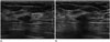
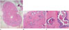
A previously healthy 38-year-old woman presented with a calcified nodule in her right breast, which was detected during routine mammography at a local clinic. She had no family history of breast cancer. On examination, no palpable lump was found in either breast or axillary region. A magnified image of the right breast revealed a 1 cm, circumscribed, isodense ovoid mass containing microcalcifications which were fine and linear in shape in the upper medial quadrant (Fig. 1). Ultrasound revealed an ovoid, well-circumscribed, parallel, hypoechoic nodule (Fig. 2). Multiple echogenic dots were present within the nodule, which correlated with the microcalcifications observed on mammography. Even though the shape, margins, and orientation of the nodule appeared benign, the microcalcifications within the mass suggested malignancy, and we diagnosed the lesion as category 4 by American College of Radiology Breast Imaging-Reporting And Data System (ACR BI-RADS) criteria. An ultrasound-guided 14-G core needle biopsy was obtained and pathology revealed atypical ductal cells in fibroadenomatous stroma. The radiologist and the pathologist agreed that the possibility of underestimation of ductal carcinoma in situ was high, and therefore, excisional biopsy was recommended. Wide excision was performed under ultrasound-guided localization of the mass. On gross examination, the cut surface of the specimen showed a hyalinized, small, round mass. Microscopic examination revealed ductal carcinoma in situ in a fibroadenoma (Fig. 3). The tumor was 0.8 × 0.6 cm in size, confined to the fibroadenoma, had a high nuclear grade with necrosis (Van Nuys group 3), and showed calcification. The immunohistochemical staining results were: ER, 3+; PR, 2+; p53, 1+; Ki-67, 2+; and CerbB-2, -. The surgical margin was free of carcinoma. The patient was given tamoxifen and has been monitored for 4 years and 4 months without any recurrence of the carcinoma.
DISCUSSION
Because carcinoma arising within a fibroadenoma is so rare, there are few reports on its radiologic features. The largest study to date was performed by Baker et al. (4), who reported the mammographic features of carcinomas originating within fibroadenomas in 24 patients. According to that study, three out of the 24 patients showed microcalcifications within the mass that suggested malignancy. On histologic examination, only one of these patients had microcalcifications associated with intraductal carcinoma in the fibroadenoma. Several cases of ductal carcinoma in situ within a fibroadenoma with suspicious microcalcifications have been reported, in which the microcalcifications were shown to be malignant (5-7). In our case, the microcalcifications within the fibroadenoma were fine and linear in shape; these types of microcalcifications are known to be correlated with a high grade of ductal carcinoma in situ (8, 9). In high-grade ductal carcinoma in situ, known as the comedo type of intraductal carcinoma, calcifications are made up of necrotic debris from dead and sloughed tumor cells within the ductal lumen.
Microcalcifications seen on mammography provide valuable information about the pathologic features of the lesion. Fibroadenoma typically involves coarse and popcorn-like calcifications during involution. According to the ACR BI-RADS, this type of calcification is classified as a typical benign finding (10), and does not warrant concern. In our case, the unusual nature of the microcalcifications within the fibroadenoma raised the possibility of malignancy and immediate pathologic confirmation was needed (10). Physicians may associate a well-defined ovoid nodule with benign fibroadenoma but should not disregard the microcalcifications within the nodule. Careful analysis and classification of microcalcifications can be very useful in making an accurate diagnosis.

 XML Download