

 PDF
PDF ePub
ePub Citation
 Print
Print


INTRODUCTION
Chronic osteomyelitis is an inflammatory disease of the bone that causes bone destruction. Malignant transformation of chronic osteomyelitis is a relatively rare but well-known complication (1). The malignant degeneration of untreated chronic wound, commonly referred to as "Marjolin ulcer" was first described in chronic burn scars in 1828 (2). There is still controversy regarding its pathophysiology; however, a latent period of 20–30 years is generally necessary for its pathogenesis (3). The plain radiographic findings are relatively well described in the literature, but reports on the magnetic resonance (MR) imaging findings are rare. We experienced a case of an 82-year-old man who was diagnosed with squamous cell carcinoma (SCC) arising from a long standing chronic osteomyelitis of the tibia.
CASE REPORT
An 82-year-old man visited our hospital with an approximately 60-year-old-history of an untreated wound in the right lower leg. He reported a laceration with bone exposure that had spontaneously healed. About 20 days prior, his right shin collided with a door. Since then, there was persistent pus-like discharge from the newly developed laceration on the chronic wound of right shin.
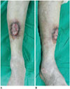
On physical examination, the patient had a wound of approximately 3 × 6 cm in size, accompanied by pus like discharge on the anterior aspect of the right shin. The wound was connected to the medullary cavity of the tibia via a draining sinus in its inferior portion. Furthermore, the skin around the wound was elevated (Fig. 1). Regional or distal lymph nodes were not palpable.
On laboratory test, the patient had an increase in erythrocyte sedimentation rate (ESR) and serum C-reactive protein (CRP) levels (ESR 54 mm/hr and CRP 3.3 mg/dL). In addition, culture tests on 2 pus samples were positive for Pseudomonas aeruginosa.
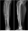
On plain radiography for an initial work-up, anteroposterior and lateral views of the right tibia showed a partially-defined heterogeneous osteolytic lesion with cortical defect on the anterior aspect of the tibial diaphysis (Fig. 2). Moreover, the anteroposterior view showed an irregular thick solid periosteal reaction.
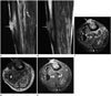
Therefore, the patient underwent MR imaging. The MR imaging revealed the presence of an ovoid shape lesion on the anterior aspect of the tibia (Fig. 3). The lesion measured approximately 5 cm in length. The lesion was heterogeneously hyper-intense as compared with the muscle on T2-weighted images and homogeneously iso- or slightly hyperintense on T1-weighted images. The lesion was also accompanied by thinning of overlying bony cortex with focal defect. Furthermore, it extended through the cortical defect and protruded into the subcutaneous layer and skin. The lesion showed heterogeneously strong enhancement on gadolinium enhancement. Adjacent bone marrow edema of approximately 10 cm length was also seen. In addition, both edema and slight contrast enhancement in the anterior and deep posterior compartment of muscles of the lower leg and the anteromedial aspect of the skin and its subcutaneous layer were observed.
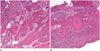
Initially, the diagnosis was granulation tissue, accompanied by the intracortical and intramedullary abscess due to chronic osteomyelitis. Therefore, the patient received saucerization and biopsy of the excised specimen. Histopathologic examination of specimens showed SCC in the skin and bone fragment (Fig. 4). Retrospective review of radiological records indicated that the lesion had both a notable contrast enhancement and an expanding appearance that distinguished it from typical granulation tissue. This led to a differential diagnosis of malignancy based on the patient's approximately 60-year-history of chronic injury.
Subsequently, the patient underwent other imaging studies, such as a positron emission tomography-computed tomography (PET-CT). The studies revealed residual SCC at the excised site, but lack of confirmed metastasis. The patient finally underwent below-knee amputation.
DISCUSSION
Malignant transformation of untreated chronic osteomyelitis is an uncommon but well-known complication, with an incidence of approximately 1.6–23% of total patients with chronic osteomyelitis (1). SCC is the most common type of malignancy, although other types of malignancy have also been described in the literature (34). Controversies exist regarding its pathophysiology; chronic purulent drainage via a draining sinus causes the degeneration and metaplasia of the skin and its subcutaneous layer (3). The latent period is reportedly longer than 10 years usually, with a mean of 30 years and maximum of 68 years (3).
Patients presenting with increased pain, foul smell, changes in drainage amount or character, mass enlargement or progressive bone destruction should be suspected for malignant transformation of chronic osteomyelitis (1). Indicated treatment is similar to that for SCC including a wide excision of the lesion or amputation (14).
The findings of plain radiography are relatively well-described in the literature. Bone destruction is the most common and important finding (14). It is commonly seen as the osteolytic or mixed sclerotic lesion (5). Moreover, it is also characterized by periosteal change and soft tissue mass or swelling (45). But these findings are not diagnostic, because these are also seen in the chronic osteomyelitis without malignant transformation (14).
MR findings of SCC arising from chronic osteomyelitis are rarely described in the literature. Soft tissue mass and bone marrow edema are the most common presentations (14). As shown in the current case, the soft tissue mass is commonly present in the skin, and may infiltrate the subcutaneous layer and invade the cortex and medulla of the underlying bony structure (67). The mass is commonly seen as hyperintense on T2-weighted images and isointense on T1-weighted images (6), but sometimes the mass is seen as hypointense on T2-weighted images due to the fibrotic nature of SCC (7). After enhancement, the mass typically shows intense enhancement, with similar enhancement of periosteum and adjacent bone cortex and medulla (89). Signal changes of bone marrow underneath the mass are typically hyperintense on T2-weighted images and hyperintense on T1-weighted images, due to preexisting chronic osteomyelitis (78). Occasionally, the sinus tract from medullary cavity to subcutaneous/cutaneous mass is found (8). MR imaging is advantageous for evaluating the extent of soft tissue mass and its invasion to the adjacent structures. But the chronic osteomyelitis also shares such characteristics as the bone marrow edema, bone destruction and soft tissue mass (14).
In this case, the patient underwent saucerization for the treatment of chronic osteomyelitis. The lesion was initially interpreted as a granulation tissue. It is difficult to differentiate between granulation tissue and malignancy, because both entities show a strong contrast enhancement (10). In the cases reported by Shimose et al. (10), elevated serum CRP levels may support the diagnosis of the osteomyelitis rather than bone tumor. In the current case, however, the patient had an increase in serum CRP levels due to the long standing chronic osteomyelitis. Further studies are therefore warranted to make a differential diagnosis between the granulation tissue and the malignant transformation of chronic osteomyelitis.
In conclusion, our case indicates that radiologists should consider the possibility of malignant transformation of chronic osteomyelitis, in patients with untreated chronic osteomyelitis presenting with bone destruction, bone marrow edema, and enhancing soft tissue mass invading bone cortex and medulla on MR imaging.
 XML Download