

 PDF
PDF ePub
ePub Citation
 Print
Print


INTRODUCTION
Gastric volvulus is defined as an abnormal degree of rotation of one part of the stomach around another (1). Gastric volvulus may lead to severe complication such as gastric ischemia, perforation, and even death. The mortality rate is reportedly 30–50% (2). Wandering spleen is defined as a movable spleen that can migrate from its normal position to any part of the abdomen or pelvis. Splenic torsion itself is a rare condition with an annual incidence of < 0.2% with or without association of wandering spleen (3). Splenic torsion is potentially fatal, if not urgently managed with surgery. It may cause ischemia, infarction, and even necrosis of the spleen (1). Gastric volvulus and wandering spleen can occur independently. However, these disease entities share a common congenital etiology of anomalous intraperitoneal visceral attachment originating from the dorsal mesogastrium. To our knowledge, only a few cases have been reported previously in literature. Herein, we described a case of gastric volvulus associated with wandering spleen and intestinal non-rotation in a 15-year-old girl, focusing on the multidetector computed tomography (MDCT) findings.
CASE REPORT
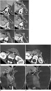
A 15-year-old girl presented at the emergency department with abdominal distention and unusual irritability. She had no underlying disease except for spastic cerebral palsy and no history of fever, trauma, toxic ingestion and previous operation. The physical examination revealed a firm, ovoid mass in the epigastric area that was non-tender. The plain radiograph of abdomen showed markedly distended stomach from diaphragm to umbilicus level. Bowel loops were unremarkable, showing no dilatation. Nasogastric tube insertion was performed for initial management. The laboratory tests were in normal range. Contrast-enhanced abdominal CT examination on a 128-detector-row CT scanner (Definition AS+, Siemens Medical Solutions, Forchheim, Germany) was performed to evaluate distended stomach. Contrast enhanced CT images demonstrated anterosuperiorly rotated gastric antrum and posteroinferiorly rotated fundus. The results indicated that the antrum was located above the gastroesophageal junction with decussate appearance, suggestive of mesenteroaxial gastric volvulus (Fig. 1A). The tip of the nasogastric tube was placed in the distal esophagus. There was no associated diaphragmatic defect. The spleen was located in the midline of supramesocolic compartment, just anterior to the pancreas that was medially displaced with kinking its tail portion (Fig. 1B-E). The splenic artery from celiac trunk was rotated in counter clockwise direction (Fig. 1D), indicating its torsion. On contrast-enhanced abdominal CT examination, the spleen showed decreased enhancement, suggesting an ischemic state. The upside-down positional change of the right gastroepiploic artery, a vascular landmark of the gastrocolic ligament, was also noted (Fig. 1E). The third and fourth portions of the duodenum and jejunal loops were located in the right side of abdomen. The patient underwent laparoscopic surgery. Surgical finding confirmed mesenteroaxial gastric volvulus. The torsion state of wandering spleen was also noted due to lack of ligamentous attachment. The gastrosplenic ligament was absent. The splenorenal ligament and the gastrocolic ligament were underdeveloped and not fixed. Duodenum and duodenojejunal junction were not fixed on the retroperitoneum. Splenopexy and gastropexy were performed. The patient recovered uneventfully and was discharged on the fifth postoperative day.
DISCUSSION
Gastric volvulus is usually divided into 2 main subtypes including organoaxial and mesenteroaxial. Organoaxial volvulus is more common than mesenteroaxial volvulus and accounts for about two-thirds of cases of gastric volvulus. Organoaxial volvulus occurs when the stomach rotates along its long axis. The greater curvature is displaced superiorly and the lesser curvature is located more caudally in the abdomen (4). In adults, this type of volvulus most commonly occurs in the setting of trauma or paraesophageal hernia and those conditions that allow the stomach to rotate along its long axis (4). On the other hand, a large Bochdalek hernia is a predisposing factor for gastric volvulus in children (4). Mesenteroaxial volvulus is much less common than organoaxial volvulus, but it is a surgical emergency and warrants prompt diagnosis and treatment. It occurs when the stomach rotates along its short axis, the transgastric axis (a line connecting the middle of the lesser curvature with the middle of the greater curvature). Mesenteroaxial volvulus results in displacement of the antrum above the gastroesophageal junction (4). Rotation is usually partial (< 180°) and is not associated with an underlying diaphragmatic defect (4). The etiology of gastric volvulus is thought to be secondary to congenital underdevelopment or acquired laxity of the supporting ligament of stomach such as the gastrohepatic, the gastrosplenic, the gastroduodenal, and the gastrophrenic ligaments, allowing approximation of cardiac and pyloric ends of the stomach, and leading to gastric volvulus (2). The unusually long gastrohepatic ligament is reportedly a predisposing factor for the development of mesenteroaxial gastric volvulus (2).
Splenic hypermobility may also be congenital or acquired (5). Congenital wandering spleen is caused by the absence or underdevelopment of splenic suspensory ligaments during fetal development. The dorsal mesogastrium fails to fuse with the posterior peritoneum, leading to absence or underdevelopment of one or all of splenic supporting ligaments such as the gastrosplenic, the splenorenal, the phrenicocolic and the phrenicosplenic ligaments (1). Among these, the spleen is anchored mainly by the gastrosplenic and the splenorenal ligaments (56). Acquired wandering spleen is caused by weakening of these ligaments due to several conditions such as hormonal effects of pregnancy, abdominal wall laxity and trauma (5). In addition, the absence of the spleen in the left upper quadrant can lead to gastric volvulus in asplenic patients (7).
In our case, the gastrosplenic ligament was absent. The splenorenal ligament and the gastrocolic ligament were underdeveloped and not fixed. These are considered as predisposing factors of coexistent gastric volvulus and torsion of wandering spleen.
Radiographic findings of gastric volvulus include a single bubble appearance of the stomach with air-fluid level. CT may show gastric volvulus and associated conditions such as perforation of the stomach, diaphragmatic defect or wandering spleen with or without complication. The CT manifestations of wandering spleen include an absence of the spleen in its normal position. It may mimic abdominal or pelvic mass. A whirled appearance of non-enhancing, twisted splenic vessels suggests acute torsion of wandering spleen. Low attenuated splenic tissue as compared with normal splenic tissue or even liver may be observed (38), as in our patient. The "rim" sign, i.e., higher density of the splenic capsule than the parenchyma, is another feature of splenic infarct and coexistent collateral circulation (5). A thick, enhancing pseudocapsule, representing omental and peritoneal adhesions may be seen in wandering spleen with chronic or intermittent torsion (8). If the pancreatic tail is involved, a whirling of pancreatic tissue and fat at the medial border of the displaced spleen may be observed on CT (8). In our case, the splenic artery and spleen partially twisted in counter clockwise direction. The pancreas was also displaced medially, and the tail portion of pancreas was kinked by the spleen, suggesting torsion of wandering spleen. The "rim" sign was not observed in our patient. Absence of the peritoneal attachment possibly hindered development of the collateral vessels supplying the spleen.
In our case, coronal reformatted CT images were helpful to trace vascular landmarks of the splenorenal ligament and the gastrocolic ligament. Upside-down positional change of the right gastroepiploic artery, a vascular landmark of the gastrocolic ligament, was observed easily on the coronal reformatted CT images. MDCT enables addition of isovoxel reconstruction images and the reformatted images facilitate the tracing of positional changes of the intraperitoneal ligaments.
Gastropexy is usually the treatment of choice for gastric volvulus. Splenopexy is also the procedure of choice to prevent future torsion when a viable wandering spleen is found at surgery (1). On the other hand, the treatment for malrotation in adults is still controversial when there is no volvulus or herniation.
In conclusion, knowledge of MDCT findings of gastric volvulus and combined wandering spleen torsion is important to make an accurate diagnosis and prevent unforeseen complications due to delayed or missed diagnosis.
 XML Download