

 PDF
PDF ePub
ePub Citation
 Print
Print


INTRODUCTION
Mesenteric fibromatosis is a rare benign fibroblastic tumor that accounts for only about 0.03% of all neoplasms and 8% of all desmoid tumors (1). Most cases of mesenteric fibromatosis occur in the small bowel mesentery (2). Most commonly, the tumor presents as a local invasion and recurrence, without metastasis. Although mesenteric fibromatosis has been reported in various sites, its presentation in the mesocolon is uncommon. Furthermore, the differential diagnosis from other mimicking tumors, such as lymphoma or adenocarcinoma in the colon, can be difficult.
In this study, we reported a case of a 47-year-old patient with mesenteric fibromatosis represented as the colo-colic type intussusception adjacent to the ascending colon that mimics malignant tumor, such as adenocarcinoma or lymphoma; furthermore, we discussed relevant radiological and pathologic findings that can be helpful for the differential diagnosis.
CASE REPORT
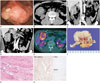
A 47-year-old male presented at our hospital with complaints of abdominal pain for 3 months prior. He had no underlying disease or any history of surgical operation. There were no specific findings on physical examination and the results of the routine laboratory test were normal. At the 2nd day of admission, he underwent the colonoscopy and there was a huge fungating mass with ulceration in the mid ascending colon. The mass was too hard for the biopsy needle to pass through easily; therefore, no specific diagnosis was made by the endoscopic biopsy (Fig. 1A). The abdomen-pelvic CT with contrast enhancement was performed on the following day for the characterization of the tumor. The pre-contrast CT scan showed a well-defined bulky mass at the right upper abdomen (Fig. 1B). This mass measured 7.2 cm in the largest diameter and revealed heterogeneous low density with no hemorrhage or calcifications. On the contrast-enhanced images, the mass presented as heterogeneous, relatively less enhanced and causing colo-colic intussusceptions (Fig. 1C, D). Colonoscopic findings of endoluminal leading point showed a lobulating contour, but an overlying inner layer suggested a well-preserved mucosa (Fig. 1C, D). In addition to the absence of bowel obstruction and other extra-intestinal manifestations, there was no pericolic fat infiltration or significantly enlarged lymph nodes adjacent to the lesion. Under the impression of a large bowel lymphoma causing colo-colic intussusception, 18F-fluorodeoxyglucose (FDG) positron emission tomography/CT (PET/CT) was performed to evaluate systemic involvement on the 4th day of admission. On the 18F-FDG PET/CT, the mass showed a high FDG uptake (maximum standardized uptake value = 5.8); other FDG uptake lesions were not detected (Fig. 1E). The preoperative impression was that of a lymphoma or an adenocarcinoma; therefore, the patient underwent laparoscopic right hemicolectomy.
Grossly, a well-defined solid mass measuring 7.5 × 7 cm abutting the ascending colon was observed. Overlying mucosa showed no abnormality. The cut surface of the mass showed a whitish color and contained whirling pattern fibers without necrosis or hemorrhage (Fig. 1F). On microscopic evaluation, the tumor invaded the muscle layer of the colonic wall. The tumor was composed of bundles of short spindle cells and collagen fiber infiltrating from serosa to the muscle layer with minimal inflammation (Fig. 1G). On immunohistochemistry stain, tumor cells were positive for beta catenin and negative for C-kit and CD-34 (Fig. 1H). Based on the immunohistochemistry results, this tumor was confirmed as mesenteric fibromatosis, benign fibroblastic proliferation.
The patients was discharged without complication and there was no recurrence during the 1 year follow-up after surgery.
DISCUSSION
Mesenteric fibromatosis is the most frequent form of intraabdominal desmoids tumor (1). Thirteen percent of patients with mesenteric fibromatosis have familial adenomatous polyposis (FAP), specifically, the Gardner syndrome variant of FAP. Prior abdominal surgery is an important risk factor in patients with FAP (2).
The CT findings of mesenteric fibromatosis can be generally categorized into the well-circumscribed form or the infiltrative form. The well-circumscribed form has a well-defined margin and shows homogeneous enhancement with similar attenuation relative to muscles (34). It is also often characterized by considerable size, which envelops the adjacent structures, especially, the small-bowel loops. In the well-circumscribed form, as in the current case, the tumor can be confused with lymphoma. The colonic lymphoma usually appears as a polypoid mass that has a circumferential wall thickening and a homogeneous enhancement (5), not very different from benign features of mesenteric fibromatosis. However, lymphoma usually has lymphadenopathy and infiltrates to other abdominal organs, such as liver and spleen, which is absent in the case of mesenteric fibromatosis (5).
The infiltrative form shows aggressive features such as an ill-defined or irregular margin and a rapid growth pattern that looks malignant (4). The imaging findings of this form are similar to those of adenocarcinoma. However, adenocarcinoma often shows metastatic lymphadenopathy and more intense contrast enhancement (6); furthermore, another key distinguishing point from mesenteric fibromatosis is the pattern in the bowel wall invasion. A desmoid tumor arises from extra-intestinal mesenteric structures such as the mesocolon, so the invasion of the muscle or the mucosal layer occurs later than in adenocarcinoma (2).
18F-FDG PET/CT is a good tool to evaluate the malignant potential of tumors based on FDG uptake (7). However, it is not the absolute cancer-specific modality (8). It is not clear why mesenteric fibromatosis could reveal false-positive imaging on 18F-FDG PET/CT like lymphoma or adenocarcinoma (78). Metser and Even-Sapir discussed many similar cases of benign lesions with high FDG uptake and their possible mechanisms. They suggested possible causes of false positive findings of 18F-FDG PET/CT like that vigorous smooth muscle activity or metabolically active mucosa (7).
In the current case, the tumor was composed of short spindle cells and collagen fiber, but there was a lack of inflammatory cells. The absence of inflammatory cells distinguishes mesen teric fibromatosis from inflammatory myofibroblastic tumor. In addition, positive immunohistochemistry finding for beta catenin supports the diagnosis of mesenteric fibromatosis (19). The high cellularity of the mass composed of fibrotic proliferation may be related to the hardened feature.
Mesenteric fibromatosis is a predisposing factor of intussusception (2). The mechanism of intussusception is believed to involve the bowel or lumen that act as irritant and provoke abnormal peristaltic movement, which may lead to the invagination of one bowel segment into the adjacent segment. Mesenteric fibromatosis also acts as lead point for an intussusception as other masses (10). Mesenteric fibromatosis can cause not only intussusception but also a small bowel obstruction or fistula formation (2). Therefore, the surgical resection is the recommended treatment option. However, the local recurrence is a major issue of mesenteric fibromatosis, even after complete surgical resection. If the resection margin of the tumor is positive or the patient shows functional loss on follow-up, an additional therapy, such as external radiation therapy, non-steroidal anti-inflammatory drug, antiestrogen therapy, or chemotherapy, can be considered (1).
In conclusion, in this study, we described a rare case of mesenteric fibromatosis involving the mesocolon, which mimicked other malignancies like lymphoma or adenocarcinoma. Such a case can be difficult to diagnose in advance of a surgical resection.
 XML Download