

 PDF
PDF ePub
ePub Citation
 Print
Print


Abstract
Purpose
The objective of this study was to review the clinical outcome after treatment of distal posterior cerebral artery (PCA) aneurysms via endovascular approach.
Materials and Methods
Eleven patients with 11 distal PCA aneurysms who were treated via endovascular approach in Inje University Busan Paik Hospital and Kosin University Gospel Hospital from December 2002 to December 2013 were retrospectively reviewed.
Results
Among the 11 patients, there were 3 males (27.3%) and 8 females (72.7%). The mean age was 56.6 years (range 44–72 years) and the mean aneurysm size was 8.45 mm (3–30 mm). Four (36.4%) aneurysms were located in the P2 segment, 6 (54.5%) in the P3 segment and 1 (9.1%) in the P1/2 junction. Seven (63.6%) aneurysms were treated with preservation of the parent artery; and the remaining 4 (36.4%) aneurysms were treated with parent artery occlusion. After treatment, the overall complication rate was 27% with the morbidity rate of 9.1% and the mortality rate of 18%.
Figures and Tables
Fig. 1
Successful embolization was done by using parent artery occlusion technique in a 59-year-old female who presented with subarachnoid and intraventricular hemorrhage (patient number 10 in Table 1).
A. Initial non-contrast enhanced computed tomography scan shows diffuse subarachnoid hemorrhage with bilateral intraventricular hemorrhage.
B, C. The anteroposterior (B) and lateral (C) projection views of a left vertebral artery angiography show a fusiform dissecting aneurysm (arrow) with ruptured state in the left P3 segment.
D. Parent artery occlusion (arrow) technique was used for complete embolization.
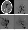
Fig. 2
A saccular dissecting aneurysm at the right P2 segment in a 61-year-old female patient who presented with headache (patient number 3 in Table 1).
A. The anteroposterior projection of right vertebral artery angiography shows a saccular dissecting aneurysm (arrow) in the right P2 segment.
B. After selective coil embolization, complete aneurysmal occlusion (arrow) was conducted without parent artery occlusion.
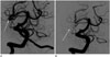
Table 1
Data for Distal PCA Aneurysm in 11 Patients
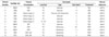
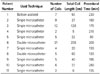
Table 2
Data for Used Technique and Materials for 11 Patients
References
1. Honda M, Tsutsumi K, Yokoyama H, Yonekura M, Nagata I. Aneurysms of the posterior cerebral artery: retrospective review of surgical treatment. Neurol Med Chir (Tokyo). 2004; 44:164–168. discussion 169
2. Chang HS, Fukushima T, Takakura K, Shimizu T. Aneurysms of the posterior cerebral artery: report of ten cases. Neurosurgery. 1986; 19:1006–1011.
3. Ciceri EF, Klucznik RP, Grossman RG, Rose JE, Mawad ME. Aneurysms of the posterior cerebral artery: classification and endovascular treatment. AJNR Am J Neuroradiol. 2001; 22:27–34.
4. Drake CG, Amacher AL. Aneurysms of the posterior cerebral artery. J Neurosurg. 1969; 30:468–474.
5. Ferrante L, Acqui M, Trilló G, Lunardi P, Fortuna A. Aneurysms of the posterior cerebral artery: do they present specific characteristics? Acta Neurochir (Wien). 1996; 138:840–852.
6. Gerber CJ, Neil-Dwyer G, Evans BT. An alternative surgical approach to aneurysms of the posterior cerebral artery. Neurosurgery. 1993; 32:928–931. discussion 931
7. Guglielmi G, Viñuela F, Duckwiler G, Dion J, Lylyk P, Berenstein A, et al. Endovascular treatment of posterior circulation aneurysms by electrothrombosis using electrically detachable coils. J Neurosurg. 1992; 77:515–524.
8. Hallacq P, Piotin M, Moret J. Endovascular occlusion of the posterior cerebral artery for the treatment of p2 segment aneurysms: retrospective review of a 10-year series. AJNR Am J Neuroradiol. 2002; 23:1128–1136.
9. Orita T, Tsurutani T, Izumihara A, Kajiwara K. Distal posterior cerebral artery aneurysms--three case reports. Neurol Med Chir (Tokyo). 1994; 34:692–696.
10. Sakata S, Fujii K, Matsushima T, Fujiwara S, Fukui M, Matsubara T, et al. Aneurysm of the posterior cerebral artery: report of eleven cases--surgical approaches and procedures. Neurosurgery. 1993; 32:163–167. discussion 167-168
11. Seoane ER, Tedeschi H, de Oliveira E, Siqueira MG, Calderón GA, Rhoton AL Jr. Management strategies for posterior cerebral artery aneurysms: a proposed new surgical classification. Acta Neurochir (Wien). 1997; 139:325–331.
12. Terasaka S, Sawamura Y, Kamiyama H, Fukushima T. Surgical approaches for the treatment of aneurysms on the P2 segment of the posterior cerebral artery. Neurosurgery. 2000; 47:359–364. discussion 364-366
13. Zeal AA, Rhoton AL Jr. Microsurgical anatomy of the posterior cerebral artery. J Neurosurg. 1978; 48:534–559.
14. Scholten FG, ter Berg HW, Hofstee N, Vellenga CJ. Giant aneurysm of the posterior cerebral artery in a one-year-old child. Eur J Radiol. 1992; 15:56–58.
15. de Sousa AA, Dantas FL, Neto AP, Carvalho GT. Giant posterior cerebral artery aneurysm in a 4-year-old child: case report. Surg Neurol. 1996; 45:31–35.
16. Lempert TE, Halbach VV, Higashida RT, Dowd CF, Urwin RW, Balousek PA, et al. Endovascular treatment of pseudoaneurysms with electrolytically detachable coils. AJNR Am J Neuroradiol. 1998; 19:907–911.
17. Lazinski D, Willinsky RA, TerBrugge K, Montanera W. Dissecting aneurysms of the posterior cerebral artery: angioarchitecture and a review of the literature. Neuroradiology. 2000; 42:128–133.
18. Pia HW, Fontana H. Aneurysms of the posterior cerebral artery. Locations and clinical pictures. Acta Neurochir (Wien). 1977; 38:13–35.
19. Sasaki O, Koizumi T, Ito Y, Sorimachi T, Koike T, Tanaka R. Dissecting aneurysm of the posterior cerebral artery treated with proximal ligation. Surg Neurol. 1992; 37:394–401.
20. Gerber CJ, Neil-Dwyer G. A review of the management of 15 cases of aneurysms of the posterior cerebral artery. Br J Neurosurg. 1992; 6:521–527.
21. Yacubian EM, Rosemberg S, da Silva HC, Jorge CL, de Oliveira E, de Assis LM. Intractable complex partial seizures associated with posterior cerebral artery giant aneurysm: a case report. Epilepsia. 1994; 35:1317–1320.
22. Kim YB, Lee JW, Huh SK, Kim BM, Kim DJ. Outcomes of multidisciplinary treatment for posterior cerebral artery aneurysms. Clin Neurol Neurosurg. 2013; 115:2062–2068.
23. Roh HG, Kim SS, Han H, Kang HS, Moon WJ, Byun HS. Endovascular treatment of posterior cerebral artery aneurysms using detachable coils. Neuroradiology. 2008; 50:237–242.
 XML Download