

 PDF
PDF ePub
ePub Citation
 Print
Print


INTRODUCTION
Computed tomography (CT) findings of thoracic abnormalities related to congestive heart failure are widely reported in the literature (1). Enlarged mediastinal lymph nodes are also known to occur in patients with congestive heart failure in several reports (23). Hence, it might be useful to identify mediastinal lymphadenopathy and other pulmonary findings associated with heart disease on CT so that invasive diagnostic approaches such as lymph node biopsy to demonstrate a benign origin can be avoided. Several previous studies have evaluated mediastinal lymph node enlargement associated with congestive heart failure, including heterogeneous disease groups such as myocardial infarction, left-sided heart failure, biventricular heart failure, etc. (24). However, in previous studies, cardiac disease-specific evaluations of mediastinal lymphadenopathy are not reported. Valvular disease is one of the most common causes of heart disease encountered in clinical practice. Despite several reports on this topic, the changes of lymph nodes in valvular heart disease have not been addressed.
The purposes of this study were to evaluate the presence, size and location of enlarged mediastinal lymph nodes in patients with valvular heart disease using CT scans and to determine the relation between lymph node enlargement and ejection fraction (EF).
MATERIALS AND METHODS
This retrospective study was approved by our Institutional Review Board.
Between April 2008 and March 2012, 98 patients with valvular heart disease who underwent CT of the chest were retrospectively identified by computerized analysis of a clinic's information system.
From this initial population, 68 patients with a pre-existing malignancy, acute infection, sarcoidosis, occupational exposure, interstitial lung disease, or a history of open cardiac surgery with potentially enlarged mediastinal lymph nodes were excluded (39 patients with suspected infection, 12 patients with interstitial lung disease, 10 patients with thoracic or extrathoracic malignancy, 6 patients undergoing valvular heart surgery, and 1 patient with pulmonary thromboembolism). Finally, the study group included 30 patients. Indications for chest CT were pre-operative evaluations for valvular surgery (21/30, 70%), dyspnea (6/30, 20%), pleural effusion (2/30, 7%), and generalized edema (1/30, 3%). We evaluated clinical follow-up using clinical information of electronic medical records to exclude the possibility of malignant or pathologic conditions of enlarged lymph nodes.
The CT examinations were obtained on multidetector CT scanners (Lightspeed VCT, GE Healthcare, Milwaukee, WI, USA, or Somatom Sensation 64 and Definition AS+, Siemens, Erlangen, Germany). Chest CT scans were obtained from the lung apices to the adrenal gland with 5-mm section thickness (n = 29) and 3-mm section thickness (n = 1). Seventeen scans were non-enhanced CT and 13 scans were contrast-enhanced CT.
The CT scans were examined separately by 2 experienced radiologists. Mediastinal lymph node enlargement was defined as short axis diameter of greater than 1 cm in the transverse plane. The size of the enlarged mediastinal lymph node was measured by 2 readers, and the average short axis diameter was determined. The locations of the enlarged mediastinal lymph nodes were recorded according to the International Association for the study of Lung Cancer. The enlarged lymph nodes were assessed for shape (oval, round) and contour (sharp, ill-defined). The presence of confluence of 2 or more enlarged lymph nodes and the presence of low attenuation with peripheral rim enhancement of lymph node were recorded.
The CT images were also analyzed for the presence of pleural effusion and pulmonary edema such as interlobular septal thickening, peribronchovascular thickening, ground-glass opacity, and increased diameter of pulmonary vessels. Other thoracic abnormalities were also recorded such as emphysema, mosaic attenuation, and etc.
In addition, the EF on transthoracic echocardiography (TTE) was reviewed for each patient using computerized analysis of clinical information. Most TTEs were performed as pre-operative evaluation for valvular surgery, and the intervals between TTE and CT were 1 day (19/30, 63%), 5 days (7/30, 23%), and 10 days (4/30, 33%).
Statistical analysis was performed with commercially available software (SPSS, version 19; IBM Corp., Armonk, NY, USA). A Comparison of frequencies was performed with the Pearson chi-squared or Fisher's exact test when tables had expected values of less than 5. A comparison between 2 groups was evaluated with the Mann-Whitney U-test. A value of p < 0.05 was considered as statistically significant.
RESULTS
This retrospective study included 30 patients (18 males and 12 females) with a mean age of 68 years (range, 36–94 years).
Valvular diseases were categorized as mitral regurgitation (MR, n = 11, 37%), aortic stenosis (AS, n = 9, 30%), aortic and mitral stenosis (AS + MS, n = 3, 10%), mitral and tricuspid regurgitations (MR + TR, n = 2, 7%), aortic regurgitation (AR, n = 2, 7%), AS and AR (AS + AR, n = 1, 3%), MS and MR (MS + MR, n = 1, 3%), and pulmonary stenosis (n = 1, 3%).
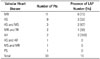
Sixteen patients (53%) had at least 1 enlarged mediastinal lymph node. Of those, 11 patients (69%) had 1 enlarged lymph node, 2 (13%) had 2 enlarged lymph nodes, 2 (13%) had 3 enlarged lymph nodes, and 1 (6%) had 5 enlarged lymph nodes. Thus, there was a total of 26 enlarged mediastinal lymph nodes. Mediastinal lymphadenopathy was seen in 8 of 11 patients with MR (73%), 3 of 9 patients with AS (33%), 2 of 3 patients with AS + MS (67%), 1 of 2 patients with MR + TR (50%), and 2 of 2 patients with AR (100%). There were no enlarged lymph nodes in patients with AS + AR, MS + MR, and PS. AR was most frequently associated with mediastinal lymphadenopathy followed by MR (Table 1).
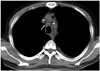
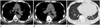
Location and mean sizes of enlarged lymph nodes were given in Table 2. The most common mediastinal lymphadenopathy locations were 4R, 4L, 7, and 2R (Figs. 1,2). The size of the enlarged lymph nodes ranged from 10.1 mm to 22.0 mm. The mean short axis diameter was 12.1 mm (standard deviation, 2.34 mm); and 25 of 26 (96%) lymph nodes were ≤ 14 mm in short axis diameter (Table 2).
Most enlarged mediastinal lymph nodes showed an oval shape (26/28 nodes, 92.9%); and only 2 lymph nodes were round (7%). All enlarged lymph nodes showed a sharp contour. A confluence of 2 or more enlarged lymph nodes was seen in 9/28 (32.1%) cases. Low attenuation with peripheral rim enhancement of lymph node was not present.
Fifteen patients had pulmonary edema, 16 patients had pleural effusions, and 13 patients had both (Figs. 1,2). Enlarged mediastinal lymph nodes were present in 12 of 15 patients with pulmonary edema (80%) and in 13 of 16 patients with pleural effusion (81%). In patients without pulmonary edema or pleural effusion (n = 12), only 2 (17%) had enlarged lymph nodes. A significantly higher frequency of mediastinal lymphadenopathy was observed in patients with pulmonary edema, pleural effusion, or both, as compared to patients without pulmonary edema or pleural effusion (p = 0.001) (Table 3).
The TTE records were reviewed for all 30 patients. The mean EF was 53.5%, ranging from 18 to 70%. In patients with mediastinal lymphadenopathy, the mean EF was 50.0%, and in patients without lymphadenopathy, it was 57.5%. Although this suggested that patients with mediastinal lymphadenopathy had a lower mean EF, there was no statistical difference in the mean EF between patients with and without mediastinal lymph node enlargement (p = 0.279). Of the 30 patients, 10 showed an abnormal EF of less than 55%. Of these, 8 patients (80%) had mediastinal lymph node enlargement, while the remaining 2 did not. However, the relationship between EF and lymph node enlargement was not statistically significant (p = 0.058) (Table 3).
Mediastinal lymph node enlargement was also studied in patients with other thoracic abnormalities including emphysema in 7 patients, mosaic attenuation in 2 patients, and arteriovenous malformation in 1 patient. Mediastinal lymph node enlargement was present in 5 of 7 patients with emphysema and in 11 of 23 patients without emphysema. There was no significant correlation between emphysema in patients with valvular heart disease and the frequency of enlarged lymph nodes (p = 0.399).
During the follow-up, 1 patient expired 1 week after operation (aortic valve replacement operation), 2 patients expired after 3 and 6 months due to congestive heart failure, and 1 patient was lost to follow-up after 5 months. Twenty-six patients were followed up for 18–89 months without evidence of malignancy.
DISCUSSION
This study was a disease-specific evaluation of the frequency of mediastinal lymphadenopathy in patients with valvular heart disease. The results suggested that valvular heart disease is often associated with mediastinal lymph node enlargement.
Enlarged mediastinal lymph nodes were observed in 16 of the 30 patients (53%) with valvular heart disease. Slanetz et al. (2) reviewed chest CT images of 46 patients with congestive heart failure. In their study, enlarged mediastinal lymph nodes were seen in 55% of cases. Chabbert et al. (5) reported 42% rate of mediastinal lymphadenopathy in 31 patients with subacute left-sided heart failure; and Erly et al. (3) reported a 66% rate of mediastinal lymphadenopathy in 44 patients with chronic cardiac dysfunction. We observed similar rates of mediastinal lymphadenopathy even though our study group included only patients with valvular heart disease.
The most frequent locations for lymph node enlargement in our study were the paratracheal and subcarinal regions, similar to the result of previous studies (25).
In our study, the size of most enlarged mediastinal lymph nodes ranged from 10 to 20 mm, which was similar to results previously reported for moderately enlarged lymph nodes (235). Slanetz et al. (2) reported that maximum short axis diameter ranged mostly between 10 to 20 mm. In the series by Erly et al. (3), 95% of the nodes were less than 20 mm in short axis diameter.
In our study, MR and AR were most frequently associated with mediastinal lymphadenopathy. Mediastinal lymphadenopathy was significantly associated with pulmonary edema and pleural effusion, and closely but not significantly related to abnormal EF. These findings suggested a relationship between mediastinal lymphadenopathy and pulmonary venous hypertension (left ventricular volume overload), which supports the pathophysiology between mediastinal abnormalities and cardiac decompression shown in several experimental investigations.
Drake et al. (6) used an experimental approach in mature sheep to show that sudden pacing-induced cardiac insufficiency was associated with a significant slowing of lymphatic flow for a pulmonary venous pressure greater than 15 cm H2O. Their data supports the fact that increases in venous pressure in heart failure interferes with lymph flow from the caudal mediastinal lymph node. When increases in pulmonary venous pressure are caused by valvular heart disease, increased pulmonary venous pressure decreases lymphatic flow from the mediastinal lymph node, resulting in mediastinal lymphadenopathy.
Unlike in other valvular diseases, the pulmonary venous hypertension induced by MR and AR is more severe due to the large amount of regurgitated blood flow and left ventricular volume overload; hence, mediastinal lymph node enlargement was more frequently noted in patients with MR and AR.
Furthermore, we observed that patients with mediastinal lymphadenopathy showed a significantly high frequency of pulmonary edema, pleural effusion, or both. This is also consistent with the fact that mediastinal lymphadenopathy in valvular heart disease is related to pulmonary venous hypertension.
Earlier studies have reported a relationship between enlarged mediastinal lymph nodes and decreased EF. Chabbert et al. (5) observed a significantly lower EF with mediastinal lymphadenopathy than without mediastinal lymphadenopathy; in addition, Erly et al. (3) observed that 81% of patients with mediastinal lymphadenopathy had an EF of less than 35%. In our study, an abnormal EF of less than 55% was frequently associated with mediastinal lymph node enlargement in patients with valvular disease. However, the relationship between EF and lymph node enlargement was not statistically significant. This difference in results is likely due to the different study populations. In our study, only patients with valvular heart disease were included, unlike other studies that included patients with congestive heart failure and various types of heart disease. EF in patients with heart failure is generally more impaired than that in patients with valvular disease. Also, we presumed that lymph node enlargement in valvular heart disease might largely depend on pulmonary venous hypertension by the large regurged flow or heart failure rather than heart failure only. The small patient population in our study could be another factor contributing to the discrepant results.
We observed that 5 of 7 patients with emphysema and 11 of 23 without emphysema showed mediastinal lymph node enlargement. Although mediastinal lymph node enlargement was more frequent in valvular heart disease patients with emphysema, this relationship was not significant in our study. However, a recent study (7) demonstrated a high percentage of enlarged hilar and mediastinal lymph nodes in patients with chronic obstructive pulmonary disease. The reason underlying the different results was likely due to the small sample size in our study.
Radiologists should be aware that mediastinal lymphadenopathy could be seen on CT in patients with non-malignant conditions such as congestive heart failure and valvular heart disease. Some studies with follow-up CT or invasive lymph node biopsy indicate that mediastinal lymphadenopathy in patients with congestive heart failure is not due to malignancy (458). Chabbert et al. (5) investigated mediastinal lymphadenopathy regression in patients with left heart failure after adequate treatment, including a systemic CT follow-up. Previously reported cases in the literature (249) also showed resolution of mediastinal lymphadenopathy and mediastinal hazy fat on follow-up CT scans. Follow-up CT scans could be useful in studying the regressive change in mediastinal lymphadenopathy associated with valvular heart disease.
Our study had some limitations. First, we included 4 patients with short term follow-up; of which 3 patients were expired due to disease related with valvular heart disease, which were an avoidable condition, and 1 patient was lost to follow-up after 5 months, which was likely too short a duration for malignancy development. Second, the size of the patient population was small. Third, due to the retrospective design of the study, different reconstruction slice thicknesses were used and contrast use in CT was inconsistent, which might have influenced the diameter measurement. Fourth, we did not demonstrate histopathological correlations for enlarged mediastinal lymph nodes.
In conclusion, the presence of mediastinal lymph node enlargement is common in patients with valvular heart disease, especially in cases of pulmonary edema and pleural effusion. Enlarged mediastinal lymph nodes were frequently observed with an abnormal EF, however, the relationship between EF and mediastinal lymphadenopathy in valvular heart disease patients was not statistically significant.


 XML Download