

 PDF
PDF ePub
ePub Citation
 Print
Print


INTRODUCTION
Abnormal insertion of the papillary muscle is a rare disorder that has been described in patients with hypertrophic cardiomyopathy (HCM). It causes left ventricular outflow tract (LVOT) obstruction and mitral regurgitation (1). However, abnormal insertion of the papillary muscle without LVOT obstruction is extremely rare with only 1 such case previously reported by Zimbarra et al. (2). Moreover, abnormal insertion of the papillary muscle was detected by echocardiography or autopsy in all studies (123), whereas, detection by computed tomography (CT) is not yet reported. Herein, we reported a case of abnormal insertion of the papillary muscle without LVOT obstruction or cardiomyopathy that was incidentally found during cardiac CT. The study was approved by the Institutional Review Board.
CASE REPORT
A 64-year-old man (height 175 cm, weight 70 kg, body surface area 1.84 m2) was admitted to our hospital for general health screening. His physical exam and results of laboratory tests were unremarkable. An electrocardiogram showed ST elevation in the V2–5 precordial leads, so cardiac CT was conducted for further evaluation of coronary artery disease. Cardiac CT scans were obtained with a 320-row detector CT scanner (Aquilion One; Toshiba Medical Systems, Otawara, Japan); scan parameters were as follows: gantry rotation time, 350 ms; tube voltage, 120 kVp; tube current, 250 mA. Nonionic contrast material (350 mgI/mL, 70 mL) was administered at 4 mL/s, followed by a 30 mL saline flush. Because his average heart rate was 70 beats per minute, CT acquisition was performed by retrospective gating with maximum exposure during the 70–80% of the RR interval [minimum exposure (100 mA tube current) in other ranges of the RR interval]. The total dose-length product was 366.4 mGy·cm and the effective dose was 5.1 mSv using a conversion factor of 0.014 mSv/mGy·cm (4). All data sets were processed with iterative reconstruction [adaptive iterative dose reduction three-dimensional (3D)] with 0.5 mm slice thickness and a 0.5 mm interval. The images were sent to the Aquarius Workstation (TeraRecon, San Mateo, CA, USA) for 3D reconstruction.
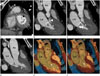
Cardiac CT showed coronary arteries arising from the coronary sinus as normal, and no evidence of plaque or luminal stenosis. On the short-axis and long-axis multiplanar reformatted images acquired during the systolic phase, the anterior papillary muscle was elongated and inserted directly into the basal portion of the anterior mitral leaflet without interposed chordae tendineae (Fig. 1A-C). However, the midcavity of the LVOT was widely opened without luminal narrowing on systolic phase (Fig. 1D). On the 3D volume-rendered long-axis views of the midcavity acquired during the diastolic phase, the anterior papillary muscle was displaced anteriorly within the left ventricular cavity. However, no evidence of LVOT obstruction at systole was found (Fig. 1E, F).
Short-axis, vertical, and horizontal long-axis cine images that were reconstructed at each 10% (10 phases) step throughout the RR interval showed normal movement of the mitral leaflet i.e., it was completely closed during the systolic phase with no definite evidence of wall motion abnormality. Short-axis images of the end-diastolic and end-systolic phases were used for global functional analysis of the left ventricle (LV). All functional parameters were normal i.e., ejection fraction, 73%; LV end-diastolic volume (EDV), 92 mL; LV end-systolic volume (ESV), 25 mL; LV EDV index, 50 mL/m2; LV ESV index, 13.6 mL/m2; cardiac output, 4.4 L/min; LV mass, 160 g; and LV mass index, 87.0 g/m2. The patient refused echocardiography for further assessment of cardiac function. All functional parameters of cardiac CT were within normal ranges and the patient had no cardiac symptoms. Therefore, follow-up echocardiography was recommended by the physician, and the patient was discharged from the hospital.
DISCUSSION
Direct insertion of the papillary muscle into the anterior mitral leaflet is a congenital malformation with a selective anomaly of chordal development at about 12 weeks' gestation (56). At this stage of normal cardiac development, the papillary muscles are contiguous with the mitral leaflets; the cephalad portions of the papillary muscles are transformed into thick muscular chordae tendineae, which ultimately become fibrous chordae positioned between the mitral leaflets and the papillary muscles (1). The selective failure of papillary muscle transformation into fibrous chordae might cause direct insertion of the papillary muscle.
Del Guzzo and Sherrid (7) described the first case of abnormal insertion of the papillary muscles with LVOT obstruction, in 1983. Subsequently, Klues et al. (1) reported anomalous papillary muscles in 10 of 78 HCM patients with marked obstruction of LVOT that appeared to be caused by systolic apposition between the hypertrophied papillary muscles and septum in the midportion of the left ventricular cavity (1). Although this anomaly is associated with HCM, it may also occur in otherwise normal hearts or in patients with other types of heart disease with or without LVOT obstruction. Recently, Zimbarra et al. (2) presented a case of anomalous insertion of the papillary muscle without LVOT obstruction in a patient with sickle cell disease; although they considered this anomaly as a normal variant, its association with sickle cell disease could not be excluded. There are no reported cases of this anomaly in healthy individuals without LVOT obstruction.
Traditionally, morphological and functional evaluation of cardiac chambers or valves is performed with echocardiography. However, echocardiography is operator-dependent and has interobserver variability; poor acoustic windows and patient's body habitus can limit image quality. In comparison, cardiac magnetic resonance imaging (MRI) provides better anatomic detail of the cardiac valves and also allows quantification of valve flow. However, cardiac MRI requires a long acquisition time and has several contraindications for certain patients. Cardiac CT is increasingly used for noninvasive coronary artery imaging due to technical developments in CT scanners. Although echocardiography remains the principal imaging technique for the assessment of the cardiac valves, cardiac CT is increasingly becoming a valuable complementary modality for visualization of the mitral and aortic valves (8). Moreover, functional analysis with cardiac CT is more accurate than that with 2-dimensional echocardiography or electrocardiography-gated single photon emission CT, which uses cardiac MRI as a reference standard (9). Therefore, an assessment of the cardiac valves and chamber anatomy during cardiac CT interpretation, whenever possible, provides valuable and detailed morphologic and functional information. Previous reports of anomalous papillary muscles are based on the echocardiographic images or autopsy. On the assumption that most of these anomalies were producing LVOT obstruction, they were detected with echocardiography in symptomatic patients.
In conclusion, we reported a rare case of incidentally detected abnormal insertion of the papillary muscle without LVOT obstruction on cardiac CT. This is the first report of the anomaly in a healthy individual who was initially assessed by cardiac CT.
 XML Download