

 PDF
PDF ePub
ePub Citation
 Print
Print


INTRODUCTION
Multiple enchondromas may be encountered in association with either enchondromatosis (Ollier's disease) or Maffucci's syndrome (1234). Although the phalanges and the metacarpals are the common sites of involvement in multiple enchondromas, secondary chondrosarcoma of the hand and feet associated with enchondromatosis is extremely rare (124). On the other hand, chondrosarcoma is a relatively common malignant bone lesion comprising approximately 10% to 12% of all malignant bone tumors; whereas, periosteal chondrosarcoma is a rare disease (25). Concurrent intramedullary and periosteal chondroid tumor in the same bone is a diagnostic challenge (6). A series of case reports on enchondromatosis provides little information on the spectrum of MR imaging findings (13). Herein, we reported a case of multiple enchondromas in a variety of locations and associated concurrent intramedullary and periosteal low grade chondrosarcomas in the lower extremities on MR imaging.
CASE REPORT
A 57-year-old female complained of pain of 6 months duration in the lateral aspect of the left foot on ambulation. Physical examination revealed a 5 × 3 cm tender, firm mass on the plantar aspect of the left mid foot. The left foot demonstrated full range of motion but mild hypesthesia of the 4th and 5th toes.
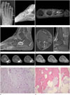
Radiographs of the left foot showed multiple osteolytic lesions with sclerotic borders in the head of the middle phalangeal head of the 2nd toe, the proximal phalangeal shaft of the 3rd toe (Fig. 1A), and the calcaneus body. Sessile exostosis-like protrusion was seen on the posterior aspect of the distal tibia. The shaft of the left 5th metatarsal bone showed subtle osteolytic lesion with sclerotic border but no cortical destruction (Fig. 1A). Radiographs of the right foot revealed intracortical chondroid tumor in the 2nd proximal phalanx and lateral aspect of the distal tibia with a similar appearance to the left.
Furthermore, MR imaging of the left foot revealed intracortical chondroid tumors in the middle phalanx of the 2nd toe and the proximal phalanx of the 3rd toe (Fig. 1B). Two calcaneal masses in the medullary space showed high signal intensity on T2 weighted image and thick rim enhancement (Fig. 1C). Concurrent eccentric chondroid lesion and sessile bony protuberance containing cartilage cap was seen on the posterolateral aspect of the distal tibia (Fig. 1D); in addition, exostosis was noted in the distal fibula.
The intramedullary lesion of the 5th metatarsal bone showed low signal intensity on T1-weighted images and high signal intensity on T2-weighted spin echo and fat suppressed T2-weighted MR images (Fig. 1E) with slight sclerotic border. The medullary lesion showed thin rim enhancement (Fig. 1F) and was 4 cm in length. However, there was no evidence of cortical destruction. A well-defined soft tissue mass was noted in the plantar aspect of the base of 5th metatarsal bone with cortical saucerization (Fig. 1EFig. 1F). There was mild soft tissue edema in the dorsum of the mass (Fig. 1E). Contrast-enhanced MR images revealed small area of nodular enhancement in the juxtacortical soft tissue mass (Fig. 1F). There was an absence of obvious linkage between the intramedullary lesion and soft tissue mass. Mild to moderate radioisotope uptake in the left 5th metatarsal bone was noted on bone scan. A preoperative diagnosis of multiple enchondromas was made based on the radiographic findings.
The medullary and cortical tumors of all the involved bones of left foot were curetted out thoroughly. Curettage with autologous bone grafting was performed in the mass of the middle phalanx of the 2nd toe, the proximal phalanx of the 3rd toe, the calcaneus body, and the 5th metatarsal bone. The soft tissue mass was excised and the root of the soft tissue mass was connected to the bone surface of the base of the 5th metatarsal bone, while maintaining the continuity of the cortex. Grossly, the mass consisted of a white, well-circumscribed, cartilaginous tumor.
Histologic examination of the resected specimen from the 2nd toe, the 3rd toe, and calcaneus revealed intracortical chondroma and enchondroma, respectively. However, histologic examination of the resected specimen from the intramedullary 5th metatarsal bone revealed grade 1 chondrosarcoma with increased cellularity within abundant myxochondroid tissue, mild to moderate nuclear atypism, and pleomorphism (Fig. 1G). Invasion of Haversian canal was noted (Fig. 1H). Histologic examination of the resected specimen of the soft tissue mass also revealed grade 1 chondrosarcoma.
The patient was discharged post-surgery without further complications. Whole body positron emission tomography-CT revealed multiple small intracortical lesions in both distal femurs with a similar appearance to the foot, in addition to both distal tibias and left distal fibula lesion. Exostosis-like lesion was also noted in the left proximal fibula. MR imaging of the right foot revealed intracortical chondroma in the 2nd proximal phalanx and eccentric chondroid tumor without cartilage cap in the lateral aspect of the distal tibia.
DISCUSSION
Enchondromatosis is a rare, nonhereditary condition that is characterized by multiple cartilaginous tumors (234). The disorder may present as asymmetric involvement of the tubular bones and pelvis. Enchondromatosis is often interchangeably termed Ollier disease. According to the original description of Ollier disease, enchondromas are predominantly confined to one side of the body and limited to the limbs. Some authors prefer the term Ollier disease for cases with extensive and unilateral distribution (4). In our case, the distribution of enchondromas was less extensive with involvement of the foot, distal tibia, and femur bilaterally.
Enchondromatosis may involve the metaphysis, diaphysis, epiphyseal plate, and articular cartilage. The enchondroma may be intracortical and periosteal (23). In our case, intracortical lesion was most common, followed by the intramedullary lesion; in addition, there was 1 periosteal lesion. Lesions that arise eccentrically within the medullary cavity simulate an osteochondroma. However, unlike osteochondroma, these protruding enchondroma have neither a cartilage cap nor a bony stalk. Our case presented with eccentric exophytic enchondroma in the left distal tibia, mimicking osteochondroma. Interestingly, the lesion fitted neither of these described conditions. Our case had cartilage cap and bony stalk on MR imaging. In addition, there was exostosis in the proximal and distal fibula. Two processes can occur simultaneously, leading to the presence of 2 lesions within the same bone (7). A Medline search resulted in many case reports of enchondromatosis. To the best of our knowledge, MR finding on protruding enchondroma with cartilage cap in the enchondromatosis has not been previously reported in the English literature. Our case adds to the spectrum of diagnostic difficulties in distinguishing enchondromatosis mimicking osteochondroma from metachondromatosis.
Malignant transformation of a solitary enchondroma to sarcoma is unusual but the risk increases in enchondromatosis by about 20–30% (1248). Most chondrosarcomas associated with enchondromatosis represent low-grade malignancy. The most difficult diagnostic problem is differentiating between enchondroma and low-grade chondrosarcoma clinically and radiographically (17). Reports on signal intensity abnormalities on precontrast and enhanced MR imaging for differentiation between enchondromas and chondrosarcomas are controversial (9). Enchondromas, as well as low-grade chondrosarcomas show septal and peripheral enhancement following contrast administration. In our case, contrast enhancement was of little value in distinguishing enchondroma from chondrosarcoma. The usual signs of malignant transformation of an enchondroma are increasing tumor size, pain of gradually increasing severity, and cortical destruction associated with soft tissue invasion (8). Approximately 2 to 3% of large (3 to 7 cm) enchondromas may undergo malignant transformation. In our case, intramedullary mass in 5th metatarsal bone was 4 cm in size. The lesion showed smooth rim enhanced chondroid tumor and lacked any radiologic sign of cortical destruction. Histologically, the differentiation of low-grade chondrosarcoma from enchondroma is also difficult. Low-grade chondrosarcoma may show similar cytological features with those of enchondroma. The growth pattern is the most important histologic feature of chondrosarcoma. Unlike enchondroma, chondrosarcomas tend to infiltrate adjacent bone marrow and Haversian canals (10). In our case, there was involvement of the Haversian canal in addition to the findings of nuclear atypism and pleomorphism.
Concurrent intramedullary and periosteal chondrosarcoma of the same bone affected with enchondroma is extremely rare and a diagnostic challenge. In the present case, chondrosarcomas developed into the medullary and the periosteal portions of the 5th metatarsal bone. In general, periosteal chondrosarcomas are of low histologic grade with invasion of the surrounding soft tissues (2510). Periosteal chondrosarcoma usually present as a painful lesion caused by stretching of the pain receptors in the periosteum (6). Periosteal chondroid tumors show soft tissue mass adjacent to the cortex, saucerization or scalloping of the adjacent cortex, and cortical sclerosis (56). Our case presented with pain and tenderness of the affected foot. MR imaging showed cortical saucerization, a well-defined soft tissue mass, and mild soft tissue edema. Differentiation of periosteal origin from intramedullary lesion with soft tissue extension might be difficult. Soft tissue involvement in the chondrosarcoma is usually associated with cortical destruction. However, in our case, cortical destruction was not detected either radiographically or grossly. Hence, it is unlikely to represent medullary chondrosarcoma with soft tissue involvement.
We observed the spectrum of less extensive distribution and various locations of enchondromatosis with associated low grade chondrosarcoma. Juxtaposed chondrosarcomas on both the medullary cavity and periosteum of the same bone add to the spectrum of diagnostic difficulties in differentiating between benign cartilage tumors and low grade chondrosarcomas. Careful clinical and radiographic evaluation of enchondromatosis is important in the diagnosis of malignant transformation of enchondroma into low grade chondrosarcoma.
 XML Download