Abstract
Eccrine poroma is a rare benign neoplasm of the eccrine sweat gland that usually presents as a small skin lesion such as a papule or nodule. This benign tumor has an overall good prognosis; however, eccrine porocarcinomas can arise from long-standing pre-existing benign eccrine poromas. We reported the case of a 37-year-old man with mental retardation who presented with an 8-cm pedunculated and densely pigmented eccrine poroma on the left hip. The tumor showed low signal intensity on T1-weighted MRI, with inhomogenously high signal intensity on T2-weighted images and strong contrast enhancement after intravenous gadolinium administration. It directly extended from the dermal layer, and the subcutaneous tissue was preserved. Radiologists should be aware that eccrine poromas could be large and pedunculated. Furthermore, related MRI findings and diagnostic clues should be carefully considered.
Eccrine poroma is a rare benign neoplasm of the acrosyringeal portion of the eccrine sweat gland (1). It may be observed on any region of the skin with eccrine glands, and usually presents as a small skin lesion primarily in the acral skin of the lower extremities (2). After complete excision, this benign tumor has a good prognosis without recurrence (3); however, eccrine porocarcinomas can arise from long-standing pre-existing benign eccrine poromas (4). Except for a few reports on eccrine porocarcinomas, MRI findings of eccrine poromas are not reported in the English literature (45). We described the MRI features of a large pedunculated and densely pigmented eccrine poroma.
A 37-year-old man presented with a 7-year history of a large mass on his left lateral hip. Given his diagnosis of mental retardation of unknown cause and moderate degree by the fourth edition of diagnostic and statistical manual of mental disorders, the precise duration of the mass was unclear. His mother detected the mass 7 years prior to presentation; however, he refused treatment and their lower socioeconomic status presented an additional barrier to care. The patient visited our outpatient clinic due to pain from the lesion.
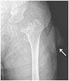
On physical examination, an 8-cm-sized, darkly pigmented soft tissue mass with a hyperkeratotic surface was noted on the skin of his left lateral hip. The lesion was soft, tender and pedunculated. No surface ulcerations, malodor, or discharge were present. On blood test and blood smear, no significant abnormality was found except macrocytic anemia due to folate and cobalamin deficiency. Plain radiography showed a large pedunculated mass in the left hip without definite intratumoral calcification. Additionally, the bilateral femoral heads were deformed due to avascular necrosis (Fig. 1). MR imaging with gadolinium enhancement was performed for further evaluation of the tumor and bilateral deformed femoral heads. On MR imaging, the soft tissue tumor of the lateral hip showed low signal intensity on T1-weighted imaging and heterogeneously mixed high and low signal intensity on T2-weighted imaging (Fig. 2A, B). The mass showed strong contrast enhancement (Fig. 2C). Dilated and tortuous vascular structures were demonstrated at the base of the tumor and underlying subcutaneous fat layer. There were mottled foci just below the surface of the periphery of the mass which showed intermediate to low signal on T1-weighted image, hyperintense on T2-weighted image and fat-suppressed T2-weighted image without contrast enhancement; hence, we regarded these as dilated glands (Fig. 2C, D). The tumor extended from the dermal layer and possessed the overlying skin layer. The possibility of tumor infiltration could not be excluded, since the subcutaneous fat tissue was grossly preserved at the base of the lesion, but the interface between the tumor and subcutaneous fat layer showed a partially indistinct margin. We regarded this mass as a malignancy originating from the dermal layer or a vascular malformation.
Skin biopsy was performed, followed by consecutive wide excision. Grossly, the excised mass was well-circumscribed with a short stalk. The outer surface of the mass was brownish tan and nodular (Fig. 3A), and the cut surface revealed a lobulated grayish white lesion involving the dermis (Fig. 3B). Histologically, it was composed of a proliferation of uniform basaloid cells with cord arrangement and interspersed ductal structures (Fig. 3C, D). The tumor cells extended from the undersurface of the epidermis in broad columns into the dermis. Focal hemorrhage and ectatic vasculature were noted in the fibrotic stroma and underlying dermis. Despite the mild cytologic atypia and frequent mitosis, it was diagnosed as a benign skin appendageal tumor with eccrine differentiation, also known as eccrine poroma.
Eccrine poroma is a rare benign neoplasm originating from the eccrine sweat gland (1). The incidence of eccrine poroma is reportedly 0.001 to 0.008% (1). It typically appears as a small nodule or papule, most commonly on the acral surface, especially the soles and lateral border of the foot. It also commonly occurs in the lower extremities and palms, fingers, neck, chest, forehead, nose and scalp; however, it can occur anywhere that eccrine glands are present (2). The etiologies and related factors of this disease remain uncertain; however, there are some reports showing an association with hormonal changes, actinic damage and trauma to the skin, immunocompromised status, chemotherapy and radiotherapy (6). It commonly occurs between 40 and 70 years of age (24), with equal prevalence in men and women (2).
Eccrine poroma is usually under 2 cm in diameter (2). Small superficial tumors can be confused with other lesions such as pyogenic granuloma, seborrheic keratosis, basal cell carcinoma and amelanotic melanoma (7). But in our case, the tumor was large, 8 × 7 × 4 cm in size, pedunculated with a thick neck, and protruded from the lateral side of the left hip. The large tumor size may have been partly due to the patient's mental retardation and late presentation to care in addition to his poor communication skills. In spite of the large size and pain, there were no features suggestive of malignancy such as growth, pruritus, bleeding or superficial ulceration (6). Reports of pigmented variants are rare (89), but our case was densely pigmented. Differential diagnosis of larger lesions includes eccrine porocarcinomas and other malignancies, including cutaneous lymphoma, squamous cell carcinoma, extramammary Paget disease, Bowen disease and cutaneous metastasis (4). Other possible differential diagnoses for the tumors originating from eccrine or apocrine gland include syringoma, hidroacanthoma simplex, dermal ductal tumor or eccrine spiradenoma.
Apart from the 2 English-language reports on the MRI findings of eccrine porocarcinomas, there are no reports on MRI findings of eccrine poromas (45). Cunningham et al. (5) reported a 1-cm-sized small foot lesion that showed slight hyperintensity on both T1- and T2-weighted MRI, as compared to the adjacent skin. Iannicelli et al. (4), reported a case with 10-cm-sized large perineal lesion that showed inhomogeneous intermediate to high signal intensity in T2-weighted images with high and non-homogeneous enhancement after gadolinium administration; however, T1 signal intensity was not reported. In our case, the extension of the tumor from the dermal layer was clear in all MR sequences (Fig. 2), confirming the dermal origin of the tumor. These MR findings have not been reported previously.
We reported on an unusual case of a benign eccrine poroma occurring at a rare site, the lateral hip. Radiologists should be aware of tumors originating from eccrine or apocrine glands, including eccrine poroma, which can be large and pedunculated. In addition, careful consideration of the MRI findings with diagnostic clues is required.
Figures and Tables
Fig. 1
Anterior-posterior radiograph shows a large pedunculated mass (arrow) in the left lateral hip. Severe osteolytic change of the left femoral head due to avascular necrosis is also seen.

Fig. 2
Axial MRI of the pedunculated skin mass occupying dermal layer in the left lateral hip.
A. On T1-weighted imaging, the mass extends from the dermal layer (arrow) and shows low signal intensity with subtle mottled low signal intensities in the periphery (arrowheads).
B. On T2-weighted imaging, the mass extends from the dermal layer (arrow) shows heterogeneously high and low signal intensity with mottled high signal intensities in the periphery (arrowheads).
C. On gadolinium-enhanced T1-weighted imaging, the mass shows strong enhancement with dilated, tortuous supplying and draining vessels in the base portion and underlying subcutaneous fat layer (arrow). The peripheral mottled lesions show low signal intensity without enhancement (arrowheads).
D. On fat-suppressed T2-weighted imaging, the mass shows heterogeneously high signal intensity. Small portion of tumor infiltration is noted at the subcutaneous fat layer (arrow). Mottled high signal intensities are observed in the periphery (arrowheads).

Fig. 3
Gross and microscopic findings with histopathologic features.
A. A dome-shaped exophytic mass with a tan brown color and surface nodularity.
B. The cut section reveals a multilobulated, tan-white, soft mass occupying the dermis layer.
C, D. Histologically, the lesion is composed of a proliferation of uniform basaloid cells with broad columns and cord arrangement with ductal structures lined by cuticles interspersed [hematoxylin-eosin stain, × 12.5 (C), × 200 (D)].

References
1. Goldman P, Pinkus H, Rogin JR. Eccrine poroma; tumors exhibiting features of the epidermal sweat duct unit. AMA Arch Derm. 1956; 74:511–521.
2. Kang MC, Kim SA, Lee KS, Cho JW. A case of an unusual eccrine poroma on the left forearm area. Ann Dermatol. 2011; 23:250–253.
3. Vu PP, Whitehead KJ, Sullivan TJ. Eccrine poroma of the eyelid. Clin Experiment Ophthalmol. 2001; 29:253–255.
4. Iannicelli E, Galluzzo A, Salvi PF, Ziparo V, David V. A large porocarcinoma of perineal region: MR findings and review of the literature. Abdom Imaging. 2008; 33:744–747.
5. Cunningham NG, Crockett RS, Kushner D. Intraepidermal eccrine adenocarcinoma of the foot: a case report. Foot Ankle Online J. 2009; [Epub]. DOI: 10.3827/faoj.2009.0207.0001.
6. Leijs MM, Merk HF, Megahed M. [Eccrine poroma]. Hautarzt. 2013; 64:328–329.
7. Horie K, Ito K, Hirata Y, Ito M. Eccrine poroma on the helix: a rare anatomical presentation. Clin Exp Dermatol. 2015; 40:442–444.
8. Allende I, Gardeazabal J, Acebo E, Díaz-Pérez JL. [Pigmented eccrine poroma]. Actas Dermosifiliogr. 2008; 99:496–498.
9. Cárdenas ML, Díaz CJ, Rueda R. Pigmented eccrine poroma in abdominal region, a rare presentation. Colomb Med (Cali). 2013; 44:115–111.


 PDF
PDF ePub
ePub Citation
 Print
Print


 XML Download