

 PDF
PDF ePub
ePub Citation
 Print
Print


INTRODUCTION
Neurofibromas are slow-growing, painless, benign nerve-sheath tumors. They occur most commonly in the dermis and subcutis, and are rarely found in the breast (1). Breast neurofibromas may develop as a result of inherited autosomal dominant neurofibromatosis type 1 (NF1) or randomly at a later time due to genetic mutations (2). They usually appear as well-circumscribed oval masses on both mammography and ultrasonography (US), and are usually located on the nipple-areolar area (234). We report a case of neurofibroma secondary to NF1 in the retromammary space resulting from endometrial cancer, which was detected on positron emission tomography-computed tomography (PET-CT) after hysterectomy.
CASE REPORT
A 50-year-old woman presented with a breast mass that showed mildly increased 18-fluorodeoxyglucose (FDG) uptake on outside PET-CT after hysterectomy for endometrioid adenocarcinoma. She had no breast-related complaints or general symptoms.
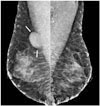
On examination, she had a 3 × 2 cm firm mass in the right upper outer (RUO) breast. She had multiple skin nodules since childhood, and her daughter and mother also had similar lesions. Mammography (Fig. 1) revealed a well-circumscribed mass measuring 32 × 19 mm at its maximum diameter, adjacent to the right pectoralis major muscle. It was located in the upper breast tissue. Multiple well-defined oval lesions with smooth and partially indistinct margins were located in both breasts. They were surrounded by lucent halos of air, indicating that they were superficial. However, the mass in the RUO breast was inconsistent with a skin lesion.
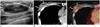
US revealed a 30 × 12 mm well-circumscribed ovoid mass with posterior acoustic enhancement in the retromammary space of the RUO quadrant, corresponding to the mass on mammography and PET-CT (Fig. 2). It abutted the pectoralis major muscle with mild compression. Five more masses in the retromammary and interpectoral areas were detected by US examination. US-guided core biopsy of a mass in the RUO quadrant was performed. The core biopsy revealed a moderately cellular spindle-cell neoplasm resembling a neurogenic tumor phyllodes tumor with stromal overgrowth and liposarcoma. An excisional biopsy was done for accurate diagnosis.
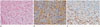
Histopathological examination of the mass showed a circumscribed, moderately cellular spindle-cell neoplasm (Fig. 3). The mass consisted of evenly distributed spindle cells with wavy nuclei. The spindle cells were intimately associated with ropy collagen bundles. Myxoid material separated the cells and collagen. Some mononuclear cells, including mast cells and lymphocytes, were identified between the cells and the collagen. No mitosis or necrosis was observed. On immunochemical staining, most cells were positive for S100 and some stromal cells were positive for CD34. A histological diagnosis of neurofibroma was made.
DISCUSSION
Neurofibromas are common benign tumors that arise from elements in the peripheral nervous system (2). They most frequently affect patients aged 20–30 years and have no sex predilection. They are commonly found as solitary lesions or are secondary to NF1, which is considered a separate disease process. When associated with NF1, most neurofibromas occur as solitary lesions in the dermis or subcutis, and multiple lesions distributed evenly over the body surface or plexiform neurofibromas characterized by diffuse neural enlargement are often found (1). Neurofibromas of the breast are quite rare in patients with NF1, and they usually occur on the nipple-areolar area (23).
Neurofibromas are considered to be benign tumors, but a small percentage, especially internal plexiform neurofibromas, have malignant cellular features that make them more likely to evolve into malignant peripheral nerve sheath tumors (MPNSTs) (5). Individuals with NF1 have a 15% increased risk of malignancy, which is a fivefold higher for breast cancer before the age of 50 years than that seen in the general population (6). Presence of multiple neurofibromas in the breast may obscure a mass in the breast at palpation, leading to delayed clinical detection (7). Although current screening guidelines do not give specific considerations, careful screening and interpretation of mammographic results for newly developing breast lesions is required.
The radiological appearance of neurofibromas varies and is known to depend on their histopathological characteristics. In the breast, neurofibromas usually appear as oval or round lesions with circumscribed margins on both mammography and US (4). On US, they appear as hypoechoic lesions with posterior acoustic enhancement, resembling a cyst, which may cause misdiagnosis. On magnetic resonance imaging, they demonstrate high signal intensity on T2-weighted images, especially if the tumor has a myxoid matrix. They may demonstrate either non-enhancement or gradual enhancement after contrast material injection (4).
In our case, a US image showed a well-circumscribed mass surrounded by extramammary fat in the retromammary area that widely abutted the pectoralis major muscle (Fig. 2A). On mammography and PET-CT, it appeared in the retromammary space and was separated from glandular tissue, indicating an extramammary location (Figs. 1, 2B). The lesion was first detected on post-operative PET-CT with mild FDG uptake. The patient had undergone a hysterectomy because of endometrioid carcinoma. Metastasis was a possible diagnosis, but most metastases of the breast are located in the parenchyma and superficially, in the fat-parenchymal interface (8). As described above, the risk of breast cancer in patients with NF1 is higher than that of the normal population (6). However, a breast cancer diagnosis was excluded because of the extramammary location of the lesion. The patient's history of NF1 and the extramammary location of the lesion were clues that led to a diagnosis of neurofibroma. MPNST, a leading cause of death in NF1 patients, cannot be reliably distinguished from neurofibroma by US (9). Therefore, if a lesion shows increased FDG uptake on PET-CT, MPNST should be considered and a histological diagnosis is necessary.
Neurofibromas are white-gray, soft, and well circumscribed but not encapsulated (10). They vary in shape and size, mostly measuring between 1 and 2 cm (45). Neurofibromas are formed by a combined proliferation of all the elements of a peripheral nerve, with Schwann cells being the most predominant entities (5). Most are immunoreactive to S100 protein and, in keeping with their benign behavior, lack significant mitotic activity and necrosis (10). Schwannomas may be differentiated from neurofibromas by the presence of Verocay bodies, Antoni A and B areas, and a more diffuse and uniform S100 staining pattern (510). These features were all absent in our case.
In conclusion, neurofibroma of the breast is rare but should be included in the differential diagnosis of breast lesion in NF1 patients. Though it can have nonspecific imaging features, its extramammary location on mammography and US is useful in distinguishing it from parenchymal lesions.
 XML Download