

 PDF
PDF ePub
ePub Citation
 Print
Print


INTRODUCTION
Meckel's diverticulum (MD) is the most common congenital anomaly of the gastrointestinal tract that occurs in 2–3% of the population (1). The MD results from incomplete obliteration of the proximal most portion of the omphalomesenteric duct within 5 to 7 weeks of fetal development (2). Gastrointestinal bleeding due to ectopic gastric mucosa of MD causing ileal peptic ulceration is the most common complication in childhood. In adults, the more common complication is bowel obstruction caused by either intussusception or small bowel volvulus around a fibrotic band or patent omphalomesenteric duct anchored to the anterior abdominal wall.
Other common complications include acute inflammation leading to perforation and hemorrhage. More rare complications include perforation of MD with foreign bodies, vesicodiverticular fistulae, strangulation in Littre's hernia, or primary neoplasms. Torsion of MD, one of the rarest complications, is the result of axial twisting around its base (3).
Pre-operative diagnosis of MD as a cause of small bowel obstruction is difficult, because the diagnosis can be made only if the diverticulum is delineated at the site of obstruction. In many cases, computed tomography (CT) features are similar to those of small bowel obstruction secondary to postoperative adhesions. Imaging features and risk factors of small bowel obstruction secondary to MD suggest the diagnosis.
Herein, we reported a rare case of MD with axial torsion; furthermore, we briefly reviewed the relevant literature.
CASE REPORT
A 21-year-old male patient presented to our emergency department with a 3 day history of cyclic crampy abdominal pain and abdominal distention. He had no known underlying disease without history of previous surgery.
Physical examination revealed normal body temperature and stable hemodynamics. The lower abdomen was distended with rebound tenderness on palpation.
Initial laboratory tests revealed a raised white blood cell count, 21.6 × 10⁹/L, and a high C-reactive protein level at 21.4 mg/dL; in addition, all other biochemical values were normal.
Initial abdominal plain radiograph showed multiple air-fluid levels in distended small bowel loops.
Initial patient management included intravenous fluid resuscitation.
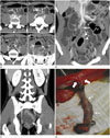
Contrast-enhanced abdominal CT examination on a 128-detector-row CT scanner (Definition AS+, Siemens Medical Solutions, Forchheim, Germany) was performed to evaluate the cause of small bowel obstruction. The CT scan showed markedly distended and fluid-loaded distal jejunal and ileal loops with bowel wall thickening. A 4.8 × 5.5 × 17.8 cm sized air and fluid containing tubular structure with distal blind end was detected in the mid-lower abdomen and pelvic cavity. An artery arising from distal ileal branch of superior mesenteric artery (SMA) was connected to the tubular structure that was suggestive of MD. The tubular structure suggestive of MD was connected to the distal ileum with a constricted neck (Fig. 1A-C). Therefore, an acute torsion of MD resulting in necrosis and perforation of MD was highly suspicious.
The patient underwent emergent laparoscopy assisted exploration. Intra-operatively, axial torsion and gangrenous diverticulum was detected at the antimesenteric border of distal ileum (about 180 cm from IC valve) (Fig. 1D). There were also a few perforated foci at the ileal loops, proximal to the diverticulum. Hence, laparoscopy assisted diverticulectomy of a 20 × 4 cm sized MD and primary repair of small bowel was performed. The patient recovered uneventfully and was discharged on the 5th postoperative day.
DISCUSSION
During the 6th week of fetal development, the midgut elongates and enters into the umbilical cord. This omphalomesenteric tract contains 3 structures that include the vitelline duct, vein, and artery. The omphalomesenteric duct divides the midgut into prearterial and postarterial limbs. The postarterial limb that is distal to the vitelline duct, elongates and becomes the distal ileum, the cecum, the ascending and the transverse colon. By the 10th week of embryogenesis, the midgut returns to the abdominal cavity and the omphalomesenteric duct becomes a thin fibrous band that is eventually absorbed (1). Incomplete absorption of the omphalomesenteric duct may result in anomalies such as umbilicoileal fistula, omphalomesenteric duct sinus, omphalomesenteric duct cyst, fibrous connection of the ileum to the umbilicus, or MD. MD is the most common (98% of cases) anomaly of the omphalomesenteric duct (1).
The MD results from complete patency of the ileal side of the duct. The diverticulum is on the antimesenteric side of the ileum and is usually found within 100 cm from the ileocecal valve. The length of postarterial limb may vary, hence, diverticula up to 180 cm are reported from the ileocecal valve (2), as in our case. MD can be subdivided by the existence of fibrous band that connects diverticulum to umbilicus. MD may connect to the umbilicus by a fibrous band if the umbilical end of the duct fails to be completely absorbed. On the other hand, MD may not be connected to the umbilicus if the umbilical end of the omphalomesenteric duct is completely absorbed (1). The fibrous band reportedly increases the chance of torsion of diverticulum (45).
The arterial blood supply and venous drainage of a MD are through remnants of the embryologic omphalomesenteric (vitellointestinal) vessels (1). Remnants and anomalies of the vitelline circulation are less common and have been reported in 8–15% of cases of MD (6). Their remnants manifest as peritoneum covered fibrous bands usually attached at the 2 extremities coursing from the ileal branch of SMA to a MD, i.e., a mesodiverticluar band, or to the anterior abdominal wall at the umbilicus (6).
On CT, MD is difficult to distinguish from normal small bowel in uncomplicated cases. However, a blind-ended fluid or gas-filled structure that is connected to ileum may be seen. On CT, small bowel obstruction secondary to MD is suspicious if a blind-ended, tubular structure that is connected with the ileum at the level of obstruction is identified. It is helpful to detect MD in the presence of vitellointestinal artery that supplies MD. The vitellointestinal artery arises from a distal ileal branch of SMA and extends to the distal ileum, which is connected to MD.
The pathogenesis of axial torsion of MD remains unclear. We reviewed the literature for possible risk factors of torsion of MD.
Several risk factors are suggested; fibrous band of remnant omphalomesenteric duct, the size and length of MD and primary neoplasm arising within MD. Fibrous band that is the partially obliterated omphalomesenteric duct, increases the chance of torsion (45). Other reported risk factors are the diverticular length and width of the diverticular sac; longer and wider diverticular sac frequently results in complications such as perforation or small bowel obstruction (578). Particularly, a narrow neck connecting the diverticulum and the ileum, increases the risk of torsion (58). In addition, primary neoplasm arising within MD is another risk factor. Although primary neoplasm of MD is rare, it is a potential risk factor. Benign as well as malignant tumors may increase the risk of torsion (5).
Diverticular torsion occurred, despite the absence of fibrous band that connects diverticulum to umbilicus in our patient. Diverticular torsion occurred because our patient had a very large diverticulum with relatively narrow neck.
In conclusion, tracing of vitellointestinal artery supplying obstructive ileal loops might suggest the diagnosis of MD in the setting of small bowel obstruction. Furthermore, blind-ended tubular structure that is connected to ileum at the level of small bowel obstruction is indicative of the diagnosis. Several factors such as a fibrous band of remnant omphalomesenteric duct, the width and length of MD with narrow neck and primary neoplasm arising within MD increase the risk of torsion.
 XML Download