

 PDF
PDF ePub
ePub Citation
 Print
Print


Abstract
Mediastinal hemangioma is a benign vascular tumor and is located most frequently in the anterior mediastinum. Computed tomography showed a well-marginated central enhancing mass with extension into the adjacent foramen. The mass was relatively hyperintense to the skeletal muscle on T2-weighted image and on fat-saturated T1-weighted image with gadolinium enhancement. The tumor was confirmed to be a cavernous hemangioma by pathologic examination after surgery. The authors recently experienced a cavernous hemangioma in the posterior mediastinum. Thus, we report a case of a posterior mediastinal mass which was difficult to differentiate from a neurogenic tumor.
Figures and Tables
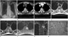
Fig. 1
A 72-year-old woman with posterior mediastinal mass.
A. Chest radiograph shows a bulging contour mass opacity (arrow) at the left paratracheal area.
B. Precontast CT of the chest shows a homogeneous mediastinal mass (mean 40 Hounsfield unit) without calcification in the posterior left paraspinal region between 3rd and 4th thoracic spine.
C. Contrast-enhanced CT of the chest demonstrates an enhancing mass similar to thoracic muscles with subtle central high density in the posterior left paraspinal region between 3rd and 4th thoracic spine.
D. Axial T1-weighted image reveals a posterior mediastinal mass on the T3 level with intermediate signal intensity similar to that of adjacent skeletal muscle and the mass extend through the intervertebral foramen.
E. Axial T2-weighted image shows a left homogeneous paraspinal mass at the same level relatively hyperintense to the skeletal muscle and the mass extend through the intervertebral foramen.
F. Axial fat-saturated enhanced T1-weighted image shows intense central enhancement of the mass at the same level and the mass extend through the intervertebral foramen.
G. Coronal fat-saturated enhanced T1-weighted image shows that a left paraspinal mass on the T3 level extend through the intervertebral foramen.
H. The lesion shows irregular shaped and varying sized cavernous vascular spaces (arrows) in the loose fibrous stroma. The vessels are thin-walled lined by single layer of endothelial cells and contains a few red blood cells in the lumen (hematoxylin-eosin stain; original magnification × 100).
References
1. Taori KB, Mitra KR, Mohite AA, Bhawatkar AJ, Siddique AQ. A case report and review of literature: posterior mediastinal haemangioma. Indian J Radiol Imaging. 2002; 12:83.
2. Yang JM, Chung WS, Kang JH, Kim YH, Kim H, Jang KS, et al. Capillary hemangioma in the posterior mediastinum: a case report. Korean J Thorac Cardiovasc Surg. 2004; 37:460–463.
3. Agarwal PP, Seely JM, Matzinger FR. Case 130: mediastinal hemangioma. Radiology. 2008; 246:634–637.
4. McAdams HP, Rosado-de-Christenson ML, Moran CA. Mediastinal hemangioma: radiographic and CT features in 14 patients. Radiology. 1994; 193:399–402.
5. Sakurai K, Hara M, Ozawa Y, Nakagawa M, Shibamoto Y. Thoracic hemangiomas: imaging via CT, MR, and PET along with pathologic correlation. J Thorac Imaging. 2008; 23:114–120.
6. Seline TH, Gross BH, Francis IR. CT and MR imaging of mediastinal hemangiomas. J Comput Assist Tomogr. 1990; 14:766–768.
7. Park HJ, Kim YT, Jou SS. Diagnostic usefulness of a multimodality study in a mediastinal hemangioma: a case report. J Korean Soc Radiol. 2010; 62:461–465.
8. Cheung YC, Ng SH, Wan YL, Tan CF, Wong HF, Ng KK. Dynamic CT features of mediastinal hemangioma: more information for evaluation. Clin Imaging. 2000; 24:276–278.
9. Ridene I, Abassi A, Baccouche I, Zidi A, Hantous-Zannad S, Marghli A, et al. Posterior mediastinal hemangioma mimicking neurogenic tumor: report of two cases. Eur J Radiol Extra. 2010; 75:e55–e58.
10. Park SJ, Cho SW, Lee HS. Venous hemangioma mimicking mediastinal solid mass-a case report. Korean J Thorac Cardiovasc Surg. 2010; 43:208–211.
 XML Download