

 PDF
PDF ePub
ePub Citation
 Print
Print


Abstract
Asbestos was previously widely used due to its many favorable characteristics, such as durability, flexibility, and inexpensiveness. Asbestos has been prohibited in Korea since 2009, however, asbestos-related diseases remain an important public health issue because of its long latency time. Lung cancer is one of the most harmful asbestos-related diseases and patients with asbestos-related lung cancer receive compensation by law. The diagnosis of asbestos-related diseases is based on a detailed interview regarding the asbestos exposure, in addition to clinical, radiological, pathological, and laboratory data. This review provides a radiologic diagnosis of asbestos-related lung cancer.
Figures and Tables
Fig. 1
Representative CT images of asbestosis; specific (A-C) and nonspecific (D-F) findings.
A. Subpleural dot-like and branching opacities.
B. Subpleural curvilinear opacities.
C. Parenchymal band.
D. Intralobular interstitial thickening (arrows).
E. Intralobular interstitial thickening (arrows) and interlobular septal thickening.
F. Honeycombing.
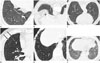
Fig. 2
Representative CT images of pleural plaque.
A. Multiple bilateral plaques.
B. Diaphragmatic pleural plaques.
C. Noncalcified plaque (arrows).
D. Thin plaque (arrows).
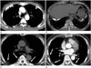
Fig. 3
A 67-year-old male who had been a slate factory worker for 20 years.
Chest PA (A) and lateral (B) views show a mass in the left middle lung zone. Pleural plaques (arrows) are seen along the right lateral chest wall and diaphragmatic pleura. Axial (C) and coronal (D) CT images show a lobulated mass with heterogeneous enhancement in left upper lobe and pleural plaques (arrows) in both hemithoraces. The mass was diagnosed as squamous cell carcinoma by percutaneous needle biopsy.
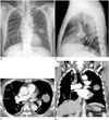
Fig. 4
A 74-year-old male who had been asbestos mine worker for 5 years and resided near the asbestos mine for 27 years.
Forty-four years after his initial exposure to asbestos, large cell neuroendocrine carcinoma developed in the right middle lobe (A). Calcified pleural plaques are seen in both hemithoraces and diaphragms (A, B). On lung window setting images (C-E), parenchymal bands and intralobular interstitial thickening suggestive of asbestosis (arrows) are noted adjacent the plaques.
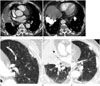
Fig. 5
A 55-year-old male with squamous cell carcinoma in the left upper lobe.
He was a construction worker for 24 years. Coronal reformatted image (A) shows a mass suggestive of lung cancer in the left upper lobe. Axial images (B, C) show honeycombing in both subpleural lungs, predominantly in the lower lung zones. In the area of less severe fibrosis, subpleural dot-like opacities, suggestive of early findings of asbestosis, are seen (arrows).
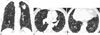
Table 1
Helsinki Criteria for Evaluating Asbestos Related Lung Cancer
Table 2
Criteria for Acceptance of Asbestos Related Lung Cancer in Asbestos Damage Relief Act in Korea
Table 3
Radiologic Diagnostic Guideline of Asbestos-Related Lung Cancer
Acknowledgments
This research was supported by the Korea Ministry of Environment under 'The Environmental Health Action program'.
References
1. Kishimoto T, Ohnishi K, Saito Y. Clinical study of asbestos-related lung cancer. Ind Health. 2003; 41:94–100.
2. Kang DM, Kim YK, Kim JE. Asbestos and environmental diseases. J Korean Med Assoc. 2012; 55:214–222.
3. Kim SG. Compensation and diagnosis of asbestos related disease. Korean J Fam Med. 2009; 30:335–343.
4. Prazakova S, Thomas PS, Sandrini A, Yates DH. Asbestos and the lung in the 21st century: an update. Clin Respir J. 2014; 8:1–10.
5. Karjalainen A, Anttila S, Heikkilä L, Kyyronen P, Vainiö H. Lobe of origin of lung cancer among asbestos-exposed patients with or without diffuse interstitial fibrosis. Scand J Work Environ Health. 1993; 19:102–107.
6. Auerbach O, Garfinkel L, Parks VR, Conston AS, Galdi VA, Joubert L. Histologic type of lung cancer and asbestos exposure. Cancer. 1984; 54:3017–3021.
7. Brodkin CA, McCullough J, Stover B, Balmes J, Hammar S, Omenn GS, et al. Lobe of origin and histologic type of lung cancer associated with asbestos exposure in the Carotene and Retinol Efficacy Trial (CARET). Am J Ind Med. 1997; 32:582–591.
8. Nielsen LS, Bælum J, Rasmussen J, Dahl S, Olsen KE, Albin M, et al. Occupational asbestos exposure and lung cancer--a systematic review of the literature. Arch Environ Occup Health. 2014; 69:191–206.
9. Roach HD, Davies GJ, Attanoos R, Crane M, Adams H, Phillips S. Asbestos: when the dust settles an imaging review of asbestos-related disease. Radiographics. 2002; 22 Spec No:S167–S184.
10. Jung JY, Ahn HS, Kim JW, Kim KA, Yun IG, Kim HW, et al. A case of asbestosis, pleural effusion and lung cancer caused by longterm occupational asbestos exposure. Tuberc Respir Dis. 1994; 41:651–657.
11. Ahn YS, Jeong KS. Epidemiologic characteristics of compensated occupational lung cancers among Korean workers. J Korean Med Sci. 2014; 29:1473–1481.
12. Ahn YS, Song JS, Kang SK, Chung HK. Understanding the occurrence of lung cancer in foundry workers through health insurance data. Korean J Prev Med. 2000; 33:299–305.
13. Yoon DY, Kang JW, Lee HJ, Kim JI, Son JE, Jung KY, et al. A case of lung cancer caused by long-term asbestos exposure. Korean J Occup Environ Med. 2004; 16:499–507.
14. Lim JW, Park SY, Choi BS. Characteristics of occupational lung cancer from 1999 to 2005. Korean J Occup Environ Med. 2010; 22:230–239.
15. Kang DM, Gu DC, Kim KH. Asbestos-related diseases among asbestos textile factory workers and residents around the factory. J Korean Med Assoc. 2009; 52:482–488.
16. Asbestos Damage Relief Center Homepage. Notice 52, Current status of asbestos damage relief judgment and certification. Incheon: Korea Environment Corporation;2015. https://www.adrc.or.kr.
17. Tossavainen A. Asbestos, asbestosis, and cancer: the Helsinki criteria for diagnosis and attribution. Scand J Work Environ Health. 1997; 23:311–316.
18. Wolff H, Vehmas T, Oksa P, Rantanen J, Vainio H. Asbestos, asbestosis, and cancer, the Helsinki criteria for diagnosis and attribution 2014: recommendations. Scand J Work Environ Health. 2015; 41:5–15.
19. Kim HR. Overview of asbestos issues in Korea. J Korean Med Sci. 2009; 24:363–367.
20. Henderson DW, Rödelsperger K, Woitowitz HJ, Leigh J. After Helsinki: a multidisciplinary review of the relationship between asbestos exposure and lung cancer, with emphasis on studies published during 1997-2004. Pathology. 2004; 36:517–550.
21. Paris C, Benichou J, Saunier F, Metayer J, Brochard P, Thiberville L, et al. Smoking status, occupational asbestos exposure and bronchial location of lung cancer. Lung Cancer. 2003; 40:17–24.
22. Lee HJ, Son JE, Hong YS, Lee YI, Yeah BJ, You CH, et al. Usefulness of high resolution computed tomography (HRCT) in the diagnosis of asbestos-related lung diseases. Korean J Occup Environ Med. 2006; 18:112–122.
23. Weiss W. Asbestosis: a marker for the increased risk of lung cancer among workers exposed to asbestos. Chest. 1999; 115:536–549.
24. Akira M, Yokoyama K, Yamamoto S, Higashihara T, Morinaga K, Kita N, et al. Early asbestosis: evaluation with high-resolution CT. Radiology. 1991; 178:409–416.
25. Kim JS. Imaging diagnosis of asbestosis. J Korean Med Assoc. 2009; 52:465–471.
26. Akira M, Yamamoto S, Inoue Y, Sakatani M. High-resolution CT of asbestosis and idiopathic pulmonary fibrosis. AJR Am J Roentgenol. 2003; 181:163–169.
27. Pairon JC, Andujar P, Rinaldo M, Ameille J, Brochard P, Chamming's S, et al. Asbestos exposure, pleural plaques, and the risk of death from lung cancer. Am J Respir Crit Care Med. 2014; 190:1413–1420.
28. Ollier M, Chamoux A, Naughton G, Pereira B, Dutheil F. Chest CT scan screening for lung cancer in asbestos occupational exposure: a systematic review and meta-analysis. Chest. 2014; 145:1339–1346.
29. National Lung Screening Trial Research Team. Aberle DR, Adams AM, Berg CD, Black WC, Clapp JD, et al. Reduced lung-cancer mortality with low-dose computed tomographic screening. N Engl J Med. 2011; 365:395–409.
30. Vehmas T, Oksa P, Kivisaari L. Lung and pleural CT signs predict deaths: 10-year follow-up after lung cancer screening of asbestos-exposed workers. Int Arch Occup Environ Health. 2012; 85:207–213.
31. Vehmas T, Oksa P. Chest HRCT signs predict deaths in long-term follow-up among asbestos exposed workers. Eur J Radiol. 2014; 83:1983–1987.
 XML Download