

 PDF
PDF ePub
ePub Citation
 Print
Print


INTRODUCTION
Multicystic dysplastic kidney is the most common form of cystic kidney disease in infants and children. It can arise from failure of the ureteric bud to integrate and branch appropriately into the metanephros during development (1). The involvement is usually unilateral, although bilateral disease can also occur, and in some patients there is segmental involvement of one kidney. Most multicystic dysplastic kidneys are benign and do not require surgical resection (2). Reported complications associated with multicystic dysplastic kidney include pain, infection, hypertension, and neoplasia (3). However, renal cell carcinomas are extremely rare in multicystic dysplastic kidneys. We found only three cases of renal cell carcinoma in previously published reports (456). Little is known regarding the imaging characteristics of this condition. We report the sonographic and CT features of a histopathologically proven cystic papillary renal cell carcinoma arising from an involutional multicystic dysplastic kidney. Institutional Review Board approval with waived informed consent (number 2014-02-009) was obtained for this case report.
CASE REPORT
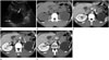
A 33-year-old male with no significant past medical history was referred for evaluation of a left retroperitoneal mass incidentally detected on ultrasonography performed at another medical institution. Findings from a physical examination were unremarkable. Laboratory examinations, including urine cytology, yielded results within normal limits. Ultrasonography of the abdomen showed a unilocular cystic mass 7 cm in diameter with multiple, mural nodules located in the left renal fossa (Fig. 1A). The left kidney was not visible. The patient underwent CT examination of the abdomen and pelvis (Somatom Sensation 16, Siemens Medical Solutions, Forchheim, Germany) before and after administration of IV contrast material (Ultravist 370, Bayer-Schering, Berlin, Germany). The mass had a primarily cystic nature with a discrete calcification in the wall which could be seen on the unenhanced image (Fig. 1B). Contrast-enhanced CT images showed peripheral soft-tissue mural nodules with mild enhancement ranging from a value of 21 Hounsfield units (HU) on the unenhanced CT image to 50 HU on the nephrographic phase obtained 90 seconds after contrast dye administration (Fig. 1C-E). There was neither lymphadenopathy nor renal vein involvement.
Because this lesion had enhancing soft-tissue mural nodules, we could not immediately rule out renal malignancy in an involutional multicystic dysplastic kidney or a cystic retroperitoneal tumor in a patient with left renal agenesis. Therefore, our patient underwent surgical exploration. Radical nephrectomy was performed without complications. Subsequent CT scanning and ultrasonography of the patient's abdomen and urine cytology were normal at a follow-up examination performed two years post-treatment.
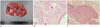
The excised cystic mass measured 8.7 × 7 × 6.5 cm. The outer surface was smooth and showed an area of fibrous thickening. On cross-section, the cystic cavity was unilocular and filled with clear, serous fluid. The inner surface showed multiple foci consisting of tan, papillary excrescences (Fig. 2A). Papillary tumor tissue, visualized microscopically, consisted of low-grade, eosinophilic renal carcinoma cells (Fig. 2B). There were also immature tubule-like structures lined by flattened or cuboidal epithelial cells scattered in the thickened areas of the wall, which suggested dysplastic kidney (Fig. 2C).
DISCUSSION
Multicystic dysplastic kidney disease is relatively common and is estimated to occur in one in 4300 live births (7). In general, patients with multicystic dysplastic kidney disease present with a unilateral abdominal mass, and as only one kidney is involved, overall renal function is normal. In general, most multicystic dysplastic kidneys spontaneously involute or decrease in size over time and have low rates of complications and malignant degeneration. Therefore, such kidneys are for the most part currently left untreated, while in the past they were routinely removed (7).
Reported complications associated with multicystic dysplastic kidney include pain, infection, hypertension, and neoplasia (6). Until now, only three reports have been published that describe renal cell carcinomas arising from multicystic dysplastic kidneys (456). In one case, the renal cell carcinoma was a papillary adenocarcinoma; histologic subtype of the other two cases is undocumented. Two of the three patients were young (15 and 26 years old). Two had mainly cystic tumors with solid components. However, these patients had aggressive tumors with early metastases involving lymph nodes, bone, and lung. Our patient is unique in that he had a cystic renal mass containing mildly enhancing mural nodules in an involutional multicystic dysplastic kidney, which replaced the normal kidney, and was asymptomatic. To our knowledge, our case is the first radiology imaging report of a renal cell carcinoma in an involutional multicystic dysplastic kidney.
Papillary renal cell carcinoma is the second most common histological subtype, accounting for 10-15% of all renal cell carcinomas (8). As a papillary renal cell carcinoma is commonly a low-stage tumor, it is associated with a very good prognosis. The most important prognostic factors for patients with renal cell carcinoma are disease stage at the time of diagnosis and nuclear grade. A tumor is more likely to be cured by surgical resection when it is small and low grade. Therefore, early detection of renal cell carcinoma is crucial in order to improve patient survival rate. Cystic papillary renal cell carcinomas, such as that in our patient, demonstrate peripheral soft-tissue mural nodules that are enhanced by administration of contrast dye. They typically appear hypovascular and homogenous on contrast-enhanced CT.
Our current understanding of multicystic dysplastic kidney is incomplete. The incidence of malignant degeneration is also unknown. The majority of multicystic dysplastic kidneys are clinically benign; some cysts spontaneously decrease in size and subsequently regress. However, as shown in previous studies, dysplastic tissue may not completely disappear despite cyst shrinkage and fluid reabsorption (6), and the malignant potential of even small amounts of residual tissue is unknown. Our case demonstrates that there is a potential risk of malignant degeneration even in an involutional multicystic dysplastic kidney. Although the incidence of malignant degeneration is low, careful consideration of elective nephrectomy is advisable in such cases.
 XML Download