

 PDF
PDF ePub
ePub Citation
 Print
Print


Abstract
Undifferentiated pleomorphic sarcoma (UPS), previously known as malignant fibrous histiocytoma, is a soft tissue sarcoma arising from mesenchymal tissue of the body. UPS of the gastrointestinal tract is known to be rare and only a few cases have been reported in the literature. Based on our case and review of the other relevant literature, the CT findings of primary UPS of the small bowel included nodular bowel wall thickening with homogeneous enhancement. It presents as a rapidly growing tumor without bowel obstruction, and it may be accompanied by distant metastasis.
Figures and Tables
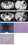 | Fig. 1Primary undifferentiated pleomorphic sarcoma (UPS) of the small bowel with adrenal metastases in a 51-year-old man.
A. The initial computed tomography (CT) image reveals segmental nodular wall thickening of the ileum with homogeneous enhancement (arrows). The degree of tumor enhancement is lower than that of normal mural enhancement. The size of the tumor is 3.6 × 2.7 cm.
B. The initial CT image presents two adrenal masses of medial (arrow) and lateral (not shown) limbs with heterogeneous enhancement. The size of upper limb tumor is 3.1 cm.
C. On a follow-up CT image 80 days later, the ileal tumor grew rapidly to 6.5 × 4.2 cm on the axial image. The tumor showing enhancing nodular wall thickening became more heterogeneous (arrows). Though the thickness of the wall is 2.0 cm or more, the patent lumen is seen as an aneurysmal dilatation.
D. On a follow-up CT image 80 days later, the right adrenal gland mass is 5.2 cm in size. It presents with central low-attenuation with a mild enhancing inner solid portion (arrow).
E. The gross pathological findings of small bowel UPS. A 6 × 5 cm tumor with diffuse necrotic changes and ulceration is noted.
F. A photomicrograph reveals the hypercellularity and storiform architecture of the pleomorphic spindle cells (hematoxylin and eosin, × 200).
G. These tumor cells are immunoreactive for alpha-1-antichymotrysin (× 200).
|
References
1. Ozzello L, Stout AP, Murray MR. Cultural characteristics of malignant histiocytomas and fibrous xanthomas. Cancer. 1963; 16:331–344.
2. Lee JH, Jee KN. Malignant fibrous histiocytoma of colon: a case report. J Korean Radiol Soc. 2006; 54:199–202.
3. Yoo DW, Shin DH, Park MS, Hur B, Lee CH. Primary malignant fibrous histiocytoma of the jejunum. J Korean Surg Soc. 2001; 60:575–578.
4. Ryu U, Lim BW, Roh JW, Lee SE, Sohn HB, Yang JH, et al. [Primary malignant fibrous histiocytoma (MFH) of the small bowel presenting as an intussusception causing small bowel obstruction]. Korean J Gastroenterol. 2004; 44:99–102.
5. Kobayashi K, Narita H, Morimoto K, Hato M, Ito A, Sugiyama K. Primary malignant fibrous histiocytoma of the ileum: report of a case. Surg Today. 2001; 31:727–731.
6. Katsourakis A, Noussios G, Hadjis I, Evangelou N, Chatzitheoklitos E. Primary malignant fibrous histiocytoma: a rare case. Case Rep Med. 2011; 2011:134801.
7. Karki B, Xu YK, Wu YK, Zhang WW. Primary malignant fibrous histiocytoma of the abdominal cavity: CT findings and pathological correlation. World J Radiol. 2012; 4:151–158.
8. Spanos C, Trigonis S, Aggelidou S, Efstratiou I, Kiskinis D, Syrrakos T. Primary malignant fibrous histiocytoma of the jejunum metastatic to the spine: report of a case. Int J Gastrointest Cancer. 2005; 35:143–145.
9. Buckley JA, Fishman EK. CT evaluation of small bowel neoplasms: spectrum of disease. Radiographics. 1998; 18:379–392.
10. Ghai S, Pattison J, Ghai S, O'Malley ME, Khalili K, Stephens M. Primary gastrointestinal lymphoma: spectrum of imaging findings with pathologic correlation. Radiographics. 2007; 27:1371–1388.
 XML Download