

 PDF
PDF ePub
ePub Citation
 Print
Print


Abstract
Small cell neuroendocrine carcinoma of the uterine cervix is a rare primary neoplasm, accounting for less than 5% of all uterine cervical cancers. The tumor is known to have an aggressive behavior and poor prognosis. In this article, we present the MRI findings of 5 cases of pathologically-proven small cell neuroendocrine carcinoma of the uterine cervix, including diffusion-weighted images.
Figures and Tables
Fig. 1
MR images (A-C), gross specimen (D), and microscopic features (E, F) of small cell neuroendocine carcinoma in a 37-year-old female (patient 5).
A. The T2-weighted sagittal image shows a 4.6 cm-sized well-marginated high signal intensity mass (arrow) on the exocervix.
B, C. The uterine cervical mass shows diffusion restriction on diffusion-weighted images (b value = 1000 sec/mm2) and the apparent diffusion coefficient value was measured to be 5.24 × 10⁻⁴ mm2/sec.
D. The gross specimen shows a 4 × 3.5 cm-sized gray-brown colored mass.
E. Microscopic findings with hematoxylin and eosin stain (× 100) include a hypercellular tumor with hyperchromatic nuclei. The tumor cells have ovoid to angulated nuclei with moulding and scanty cytoplasm. Abundant apoptotic activities and mitoses are present.
F. An immunohistochemical stain of synaptophysin (× 100) shows diffuse strong positivity in tumor cells.
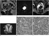
Table 1
Clinical and Image Findings of 5 Patients with Small Cell Neuroendocrine Carcinoma in Uterine Cervix
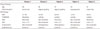
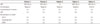
Table 2
Histologic Findings of 5 Patients with Small Cell Neuroendocrine Carcinoma in Uterine Cervix
References
1. Klimstra DS, Modlin IR, Coppola D, Lloyd RV, Suster S. The pathologic classification of neuroendocrine tumors: a review of nomenclature, grading, and staging systems. Pancreas. 2010; 39:707–712.
2. Viswanathan AN, Deavers MT, Jhingran A, Ramirez PT, Levenback C, Eifel PJ. Small cell neuroendocrine carcinoma of the cervix: outcome and patterns of recurrence. Gynecol Oncol. 2004; 93:27–33.
3. Abeler VM, Holm R, Nesland JM, Kjørstad KE. Small cell carcinoma of the cervix. A clinicopathologic study of 26 patients. Cancer. 1994; 73:672–667.
4. Kim do Y, Yun HJ, Lee YS, Lee HN, Kim CJ. Small cell neuroendocrine carcinoma of the uterine cervix presenting with syndrome of inappropriate antidiuretic hormone secretion. Obstet Gynecol Sci. 2013; 56:420–425.
5. Yang DH, Kim JK, Kim KW, Bae SJ, Kim KH, Cho KS. MRI of small cell carcinoma of the uterine cervix with pathologic correlation. AJR Am J Roentgenol. 2004; 182:1255–1258.
6. van Nagell JR Jr, Powell DE, Gallion HH, Elliott DG, Donaldson ES, Carpenter AE, et al. Small cell carcinoma of the uterine cervix. Cancer. 1988; 62:1586–1593.
7. Rashed MM, Bekele A. Neuroendocrine differentiation in a case of cervical cancer. Pan Afr Med J. 2010; 6:4.
8. Kuang F, Ren J, Zhong Q, Liyuan F, Huan Y, Chen Z. The value of apparent diffusion coefficient in the assessment of cervical cancer. Eur Radiol. 2013; 23:1050–1058.
 XML Download