

 PDF
PDF ePub
ePub Citation
 Print
Print


INTRODUCTION
Right-sided infective endocarditis (RSIE) accounts for only 5-10% of cases of infective endocarditis (IE), the majority of which involve the tricuspid valve (TV) and occur predominantly in intravenous drug abusers, patients with pacemakers or central venous lines, or those with congenital heart disease (1). Diagnosis of IE is based on the modified Duke criteria including clinical, biological and imaging findings. Echocardiography is usually the first-line diagnostic study performed. According to modified Duke criteria, positive echocardiographic findings for IE include an oscillating intracardiac mass on a valve or supporting structure, in the path of regurgitant jets, or on implanted material in the absence of an alternative anatomical explanation; or abscess or new partial dehiscence of a prosthetic valve (2). Multidetector computed tomography (CT) also may be useful for identifying valvular vegetation or abscess, which may support diagnosis of IE (3). Here, we describe a case of RSIE with tumor-mimicking right ventricular vegetation in a patient with no history of congenital heart disease or intravenous drug abuse, which was documented on both echocardiography and cardiac CT.
CASE REPORT
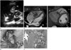
A 45-year-old man was transferred to our institution following a 2-week bout of general weakness and uncontrolled fever despite antibiotic use at an outside hospital. The patient denied any history of heavy alcohol intake, drug abuse, or structural heart disease. Physical examination was unremarkable, except that the patient had a body temperature of 38.9℃. His electrocardiogram was normal. Laboratory results showed an elevated white blood cell count (18400/mm3; normal range, 4000-10000/mm3), C-reactive protein level (104 mg/L; normal range, 0-0.3 mg/L), and erythrocyte sedimentation rate (98 mm/hour; normal range, 0-22 mm/hour). Blood cultures were positive for Streptococcus viridans (S. viridans). Transthoracic echocardiography (TTE) revealed a 4.5 × 2.3 cm highly mobile homogeneous mass (arrow) attached to the TV (arrowhead), along with a moderate degree of tricuspid regurgitation (Fig. 1A). On CT images of the right ventricle adjacent to the TV, the lesion presented as a 4 cm irregular low-density mass (arrows) without any evidence of calcification (Fig. 1B, C). The attenuation value was 56 Hounsfield units (HU) on a precontrast scan and 75 HU on an arterialphase postcontrast scan. The mass exhibited mild homogeneous enhancement after contrast medium injection. On virtual endoscopic (Fig. 1D) and 4-chamber (Fig. 1B) CT reconstructed images, the mass seemed to show attachment to the TV with a stalk (arrow in Fig. 1D, arrowhead in Fig. 1B). Chest radiography revealed nodular opacity in the right lower lobe, while contrast-enhanced electrocardiography-gated 64-slice cardiac CT (Discovery 750 HD; General Electric Medical Systems, Milwaukee, WI, USA) showed poorly defined nodules along the bronchovascular bundles and in the subpleural areas of both lungs, consistent with septic lung. Additionally, on CT images, a filling defect was noted in the superior segmental branch of the right lower pulmonary artery, suggesting the possibility of septic embolism. As a differential diagnosis for the mass in the right ventricle, considering the patient's symptoms and signs including fever andpositive blood culture results for S. viridans which suggests possible IE, vegetation and thrombus and tumorous conditions such as myxoma or papillary fibroelastoma were considered.
During surgery to remove the mass, a 4 cm entity was seen that was attached to the anterior and septal leaflet of the TV. Pathologic examination of the resected specimen from the right ventricle revealed multiple bacterial colonies intermingled with fibrinopurulent inflammatory exudate, consistent with vegetation (Fig. 1E). Finally, according to the modified Duke criteria (2), definite diagnosis of IE was established. Following surgery, the patient underwent 4 weeks of antimicrobial therapy, after which his symptoms were relieved and follow-up TTE showed no evidence of further pathology.
DISCUSSION
The majority (73%) of patients with RSIE have pre-existing congenital heart disease or an acquired valvular lesion; cases of RSIE without any predisposing factors are relatively rare. Our patient represents the subset of individuals without a history of drug use or any evidence of underlying cardiac abnormality (4).
Guidelines consider TTE to be the gold standard imaging modality for suspected IE. However, it is difficult to differentiate between vegetation, calcification, and tumor using echocardiography, because all three conditions appear hyperechogenic. Furthermore, echocardiography requires a highly trained technician, and results are technician-dependent. On CT, however, calcification will appear hyperdense, whereas soft tissue masses will appear hypodense. Contrast-enhanced cardiac CT may be helpful in further differentiating between vegetation, thrombus, and tumor. However, at times it may be impossible to differentiate between a small mobile thrombus from vegetation or a large vegetation from a tumor. Nonetheless, cardiac CT has shown promising results with regard to valvular and perivalvular damage by providing high-resolution anatomical information and affording multiplanar reformations (5).
In our patient, tumor-mimicking vegetation was noted as a large mobile homogeneous mass on echocardiography and as a noncalcified mildly enhanced mass in the right ventricle on cardiac CT. Vegetation, tumor such as myxoma, and thrombus were considered in the differential diagnosis. Cardiac papillary fibroelastomas are also well-known tumors that usually arise from the valvular endocardium, though they generally present as small mobile pedunculated or sessile valvular or endocardial masses on echocardiography (6). Cardiac myxomas predominantly occur in the left atrium, whereas valvular myxomas are extremely rare. Echocardiographic characteristics of valvular myxomas include a globular shape and a heterogeneous or mottled appearance throughout, with echolucent areas representing hemorrhage or necrosis (7). On CT images, myxomas often present as hypodense or isodense with respect to the myocardium, and may accompany coarse dystrophic calcification. On contrast-enhanced cardiac CT, myxomas appear as intracavity filling defects with heterogeneous contrast enhancement, though attenuation may be variable depending on chronicity and whether necrosis or hemorrhage are present. In our patient, the uncontrolled fever, positive blood culture results for S. viridans, and final pathologic confirmation of the lesion as vegetation established the definite diagnosis of IE. Moreover, the findings suggesting septic lung and septic embolism of the branch of the right pulmonary artery on CT supported such a diagnosis. Although there are no definitive indications of IE that can be seen on CT scans, according to Grob et al. (8), the key findings are vegetation, perforation of the valve, valvular aneurysm and perivalvular abscess. Valvular vegetation can appear as a thickened valve or as an irregular homogeneous hypodense mass attached to the valve or other endocardial structure, which is usually mobile during the cardiac cycle.
Prognosis following a diagnosis of RSIE is relatively good, with an in-hospital mortality rate of < 10%. According to a study by Martín-Dávila et al. (9), vegetation length > 20 mm and fungal etiology were the main predictors of death in cases of RSIE in intravenous drug abusers. RSIE is resolved with non-invasive procedures in 70-85% of cases, with the remaining cases requiring surgical management. Surgical treatment should be considered in the following situations: right heart failure secondary to severe tricuspid regurgitation with poor response to diuretic therapy, IE caused by an organism that is difficult to eradicate (e.g., persistent fungi) or bacteremia that persists for at least 7 days (e.g., that caused by Staphylococcus aureus, or Pseudomonas aeruginosa) despite adequate antimicrobial therapy, and TV vegetation > 20 mm that persists after recurrent pulmonary emboli with or without concomitant right heart failure (10). Surgery was performed appropriately in our patient as his symptoms were relieved by the treatment.
In sum, we report the echocardiographic and CT findings of large right ventricular vegetation, which was pathologically confirmed in a patient with IE.
 XML Download