

 PDF
PDF ePub
ePub Citation
 Print
Print


Abstract
Purpose
We aimed to evaluate the utility of two types of MR cisternography [fast spin echo sequence and steady-state coherent gradient echo (GRE) sequence] in addition to phase contrast-cine imaging (PC-cine), for assessing patency at the aqueduct and third ventriculostomy site.
Materials and Methods
43 patients (35 patients with suspected aqueductal stenosis and 8 patients with third ventriculostomy) were retrospectively analyzed. PC-cine, 3 dimensional sagittal fast spin echo sequence [driven-equilibrium imaging (DRIVE) or volumetric isotrophic T2-weighted acquisition (T2 VISTA)] and steady-state coherent fast GRE sequence (balanced turbo field echo; bTFE) imaging were performed in all patients. The patency of the aqueduct or third ventriculostomy site was scored. Some pitfalls of each sequence were also analyzed in individual cases.
Results
93% of all cases showed consistent scores in PC-cine, DRIVE/T2 VISTA, and bTFE imaging. DRIVE/T2 VISTA imaging provided functional information of cerebrospinal fluid flow with flow-related artifacts, while bTFE imaging allowed direct visualization of the aqueduct or ventriculostomy site. However, evaluation of anatomical structures was difficult in three cases with strong flow-related artifacts on DRIVE/T2 VISTA and in 2 cases with susceptibility artifacts on bTFE.
Figures and Tables
Fig. 1
A 47-year-old man with Chiari malformation.
A. Phase-contrast cine image shows active cerebrospinal fluid pulsation through the aqueduct (arrows).
B. Volumetric isotrophic T2-weighted acquisition image shows patent aqueduct and flow-related artifact within the patent aqueduct (arrows).
C. Balanced turbo field echo image shows patent aqueduct without flow-related artifact (arrow).
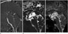
Fig. 2
A 54-year-old man with aqueductal stenosis.
A. Phase-contrast cine image shows faint cerebrospinal fluid pulsation through the aqueduct (arrows).
B, C. Both volumetric isotrophic T2-weighted acquisition (T2 VISTA) (B) and balanced turbo field echo (C) images show web-like lesion at the superior medullary velum of the 4th ventricle causing aqueductal stenosis (arrow). Also there is no flow-related artifact through the aqueduct on T2 VISTA image (B).
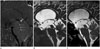
Fig. 3
A 65-year-old woman with a coil-embolized aneurysm at the top of the basilar artery. Phase-contrast cine image (A) shows coil-embolized giant aneurysm at the basilar tip (arrowheads) and no active cerebrospinal fluid pulsation through the aqueduct (arrow). Volumetric isotrophic T2-weighted acquisition image (B) also shows coil-embolized aneurysm causing aqueductal stenosis (arrow). However, the lumen of the aqueduct is obscured by coil-induced susceptibility artifact on balanced turbo field echo image (C).
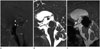
Fig. 4
A 3-year-old boy with Dandy-Walker variant. Phase-contrast cine image (A) shows active cerebrospinal fluid (CSF) pulsation through the aqueduct (arrow). Volumetric isotrophic T2-weighted acquisition image (B) shows severe flow-related artifact through the aqueduct due to large amount of CSF flow, which obscures the normal contour of the aqueduct (arrow). Balanced turbo field echo image (C) shows slightly widened aqueduct and minimal flow-related artifact within the aqueduct (arrow).

Fig. 5
A 23-year-old man underwent the endoscopic third ventriculostomy for the aqueductal stenosis. Phase-contrast cine image (A) shows active cerebrospinal fluid pulsation through the ventriculostomy site in the 3rd ventricle floor (arrows). Volumetric isotrophic T2-weighted acquisition image (B) directly demonstrates the ventriculostomy site (arrows) and flow-related artifact through the ventriculostomy site. The ventriculostomy site is directly identified without flow-related artifact on balanced turbo field echo image (arrows) (C).
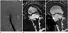
Fig. 6
A 10-month-old girl with endoscopic third ventriculostomy for the aqueductal stenosis. Phase-contrast cine image (A) shows active cerebrospinal fluid pulsation through the ventriculostomy site (arrow). Driven-equilibrium image (B) shows severe flow-related artifact in the 3rd ventricle and prepontine cistern area (arrows), which obscures the exact ventriculostomy site at the 3rd ventricle floor, whereas 3rd ventriculostomy site is localized with minimal linear flow-related artifact on balanced turbo field echo image (arrows) (C).
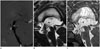
Table 1
Comparison of PC-Cine, T2 VISTA/DRIVE and bTFE Imaging Scores for Evaluation of Aqueductal Patency
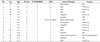
*Y = years, M = months.
AC = arachnoid cyst, bTFE = balanced turbo field echo, DRIVE = driven-equilibrium imaging, ETV = endoscopic third ventriculostomy, NA = not assessed, PC-cine = phase contrast-cine imaging, T2 VISTA = volumetric isotrophic T2-weighted acquisition, VPS = ventriculoperitoneal shunt
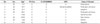
Table 2
Comparison of PC-Cine, T2 VISTA/DRIVE and bTFE Imaging Scores for Evaluation of Patency at Ventriculostomy Site after Endoscopic Third Ventriculostomy
References
1. Tisell M. How should primary aqueductal stenosis in adults be treated? A review. Acta Neurol Scand. 2005; 111:145–153.
2. Kunz M, Schulte-Altedorneburg G, Uhl E, Schmid-Elsaesser R, Schöller K, Zausinger S. Three-dimensional constructive interference in steady-state magnetic resonance imaging in obstructive hydrocephalus: relevance for endoscopic third ventriculostomy and clinical results. J Neurosurg. 2008; 109:931–938.
3. Dinçer A, Yildiz E, Kohan S, Memet Özek M. Analysis of endoscopic third ventriculostomy patency by MRI: value of different pulse sequences, the sequence parameters, and the imaging planes for investigation of flow void. Childs Nerv Syst. 2011; 27:127–135.
4. Algin O, Hakyemez B, Parlak M. Phase-contrast MRI and 3D-CISS versus contrast-enhanced MR cisternography on the evaluation of the aqueductal stenosis. Neuroradiology. 2010; 52:99–108.
5. Kurihara N, Takahashi S, Tamura H, Higano S, Furuta S, Jokura H, et al. Investigation of hydrocephalus with three-dimensional constructive interference in steady state MRI. Neuroradiology. 2000; 42:634–638.
6. Algin O, Turkbey B. Evaluation of aqueductal stenosis by 3D sampling perfection with application-optimized contrasts using different flip angle evolutions sequence: preliminary results with 3T MR imaging. AJNR Am J Neuroradiol. 2012; 33:740–746.
7. Algin O, Turkbey B, Ozmen E, Ocakoglu G, Karaoglanoglu M, Arslan H. Evaluation of spontaneous third ventriculostomy by three-dimensional sampling perfection with application-optimized contrasts using different flip-angle evolutions (3D-SPACE) sequence by 3T MR imaging: preliminary results with variant flip-angle mode. J Neuroradiol. 2013; 40:11–18.
8. Stoquart-El Sankari S, Lehmann P, Gondry-Jouet C, Fichten A, Godefroy O, Meyer ME, et al. Phase-contrast MR imaging support for the diagnosis of aqueductal stenosis. AJNR Am J Neuroradiol. 2009; 30:209–214.
9. Jung NY, Moon WJ, Lee MH, Chung EC. Magnetic resonance cisternography: comparison between 3-dimensional driven equilibrium with sensitivity encoding and 3-dimensional balanced fast-field echo sequences with sensitivity encoding. J Comput Assist Tomogr. 2007; 31:588–591.
10. Kwon JW, Yoon YC, Choi SH. Three-dimensional isotropic T2-weighted cervical MRI at 3T: comparison with two-dimensional T2-weighted sequences. Clin Radiol. 2012; 67:106–113.
11. Tsuchiya K, Aoki C, Hachiya J. Evaluation of MR cisternography of the cerebellopontine angle using a balanced fast-field-echo sequence: preliminary findings. Eur Radiol. 2004; 14:239–242.
 XML Download