

 PDF
PDF ePub
ePub Citation
 Print
Print


Abstract
The spontaneous infarction of benign breast lesions is a rare entity and hence is not usually considered in the differential diagnosis during radiologic or clinical examination. There have been a few published cases of infarction during pregnancy and lactation. In this study we report the ultrasonographic and pathologic features of a spontaneous infarction of a lactating adenoma with acute mastitis and abscess and a spontaneously infarcted fibroadenoma.
Figures and Tables
Fig. 1
A 30-year-old woman with an edematous change in the left breast 5 days after delivery.
A. Mediolateral oblique mammogram of the left breast shows a large circumscribed heterogeneously hyperdense mass, replacing the whole left breast. The homogeneously hyperdense portion (star) in the periphery of the mass correlates with the outer portion of the surgical specimen, suggesting lactating adenoma without infarction. Furthermore, the irregular central portion with air densities (asterisk) correlates with the specimen, and suggesting infarction with necrosis.
B. Ultrasonography shows a heterogeneously hypoechoic mass. Internal multiple echogenic foci with posterior shadowing (asterisk) correlates with the infarcted portion of the lactating adenoma. Skin thickening (arrow) suggests combined acute inflammatory change.
C. Color Doppler image shows the lack of internal vascularity.
D. Macroscopic view of the excised specimen shows a large lactating adenoma (star) with an internal necrotic portion (asterisk).
E. Photomicrograph of the specimen shows features of an infarcted lactating adenoma. Extensive coagulative necrosis and the lack of tissue staining with preserved reticulin architecture suggest infarction. Moreover, vascular thrombosis (arrow) is also detected (hematoxylin and eosin, × 100 magnification in the main figure and × 200 magnification in the inset figure).
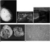
Fig. 2
A 24-year-old pregnant woman (32 weeks) with a palpable mass in her right breast.
A. Ultrasonography shows a circumscribed complex solid and cystic mass with internal multiple small anechoic portions (asterisks).
B. Photomicrograph of the specimen shows features of an infarcted fibroadenoma. Compared with residual viable tissue (left, star), extensive coagulative necrosis and the lack of tissue staining with preserved reticulin architecture (right) suggest infarction (hematoxylin and eosin, × 200).
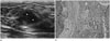
Table 1
Ultrasonographic Features of Previously Reported Cases of Infarction in Benign Breast Lesions
| Year | Authors | Age (Years) | Sex | Breast Lesion | Ultrasonographic Features |
|---|---|---|---|---|---|
| 1999 | Behrndt et al. (1) | 31, pregnant at 35 weeks | F | Lactating adenoma | Circumscribed heterogeneous hypoechoic lesion |
| 2004 | Fowler (10) | 12 | F | FA | Complex solid and cystic lesion |
| 2005 | Hsu et al. (8) | 12 | F | FA | Circumscribed homogeneous hypoechoic lesion |
| 2007 | Sabate et al. (2) | 35, 4 months after delivery | F | FA | Not circumscribed heterogeneous hypoechoic lesion with acoustic enhancement |
| 2009 | Oh et al. (4) | 31, 3 months after abortion | F | FA | Circumscribed heterogeneous hypoechoic lesion with internal anechoic portion and acoustic enhancement |
| 2013 | Skenderi et al. (3) | 13 | F | Juvenile FA | Circumscribed homogeneous hypoechoic lesion |
| 2014 | Kim (7) | 15 | F | FA | Complex solid and cystic lesion with acoustic enhancement |
References
1. Behrndt VS, Barbakoff D, Askin FB, Brem RF. Infarcted lactating adenoma presenting as a rapidly enlarging breast mass. AJR Am J Roentgenol. 1999; 173:933–935.
2. Sabate JM, Clotet M, Torrubia S, Gomez A, Guerrero R, de las Heras P, et al. Radiologic evaluation of breast disorders related to pregnancy and lactation. Radiographics. 2007; 27:Suppl 1. S101–S124.
3. Skenderi F, Krakonja F, Vranic S. Infarcted fibroadenoma of the breast: report of two new cases with review of the literature. Diagn Pathol. 2013; 8:38.
4. Oh YJ, Choi SH, Chung SY, Yang I, Woo JY, Lee MJ. Spontaneously infarcted fibroadenoma mimicking breast cancer. J Ultrasound Med. 2009; 28:1421–1423.
5. Lucey JJ. Spontaneous infarction of the breast. J Clin Pathol. 1975; 28:937–943.
6. Oh HJ, Kim SH, Kang BJ, Lee AW, Song BJ, Kim HS, et al. Ultrasonographic features of spontaneous breast tumor infarction. Breast Cancer. 2014; Mar. 15. [Epub]. DOI: 10.1007/s12282-014-0525-3.
7. Kim SJ. Spontaneously infarcted fibroadenoma of the breast in an adolescent girl: sonographic findings. J Med Ultrasonics. 2014; 41:83–85.
8. Hsu S, Hsieh H, Hsu G, Lee H, Chen K, Yu J. Spontaneous infarction of a fibroadenoma of the breast in a 12-year-old girl. J Med Sci Taipei. 2005; 25:313.
9. Chang YW, Kwon KH, Goo DE, Choi DL, Lee HK, Yang SB. Sonographic differentiation of benign and malignant cystic lesions of the breast. J Ultrasound Med. 2007; 26:47–53.
10. Fowler CL. Spontaneous infarction of fibroadenoma in an adolescent girl. Pediatr Radiol. 2004; 34:988–990.
 XML Download