

 PDF
PDF ePub
ePub Citation
 Print
Print


INTRODUCTION
The tensor fasciae suralis muscle, also known as the ischioaponeuroticus, is a rare accessory muscle. It may originate from both of the semitendinosus and the long head of the biceps femoris, or from either of them, and inserts into the sural fasciae or the Achilles tendon (1). Although it may present as a popliteal mass, it may also be detected incidentally (23).
We encountered this anatomic variation in the popliteal region, as an incidental finding on magnetic resonance imaging (MRI). The following ultrasound (US) correlated well with the MRI findings, confirming the diagnosis.
CASE REPORT
A 30-year-old man visited the hospital for right knee pain following an injury after playing basket ball. He denied previous local injury or surgery.
Physical examination revealed tenderness, swelling and range-of-motion limitation in the right knee joint. X-ray showed joint effusion without bony abnormality.
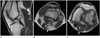
MRI of right knee was performed using a 1.5-T MR scanner (Signa HDxt 1.5T; GE Medical Systems, Milwaukee, WI, USA). The examination revealed the tensor fasciae suralis muscle arising from the lateral aspect of semitendinosus muscle, running superficial to the gastrocnemius medial head (Fig. 1). The other abnormality was complete tear of the anterior cruciate ligament.
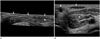
Subsequently, sonography was carried out with the patient prone using a linear 12-5 MHz array transducer (iU22; Philips Medical Systems, Bothell, WA, USA). The sonograms showed the tensor fasciae suralis muscle as an elongated structure with hyperechoic striations on a hypoechoic background, which is typical echotexture of the normal muscle in the right popliteal region. The muscle extended inferiorly and was attached to the gastrocnemius medial head (Fig. 2).
The patient underwent anterior cruciate ligament reconstruction. His pain disappeared after operation and he was followed up periodically as an outpatient.
DISCUSSION
There are several known accessory muscles of the knee. Accessory slips of the medial or lateral gastrocnemius muscle are frequently encountered in popliteal area on knee MRI. An anomalous relationship between the popliteal artery and the proximal gastrocnemius may manifest clinically with popliteal artery entrapment syndrome (4). The variant muscle in superficial popliteal fossa may simulate soft tissue tumors (5). Of these, the tensor fasciae suralis is located superficially in the popliteal fossa, between the semitendinosus and semimembranosus muscles medially and the biceps femoris muscle laterally (4). Although it may arise from the distal aspect of any of the hamstring muscles, the majority of reported cases indicated that it originates from the distal semitendinosus muscle. It may insert into the posterior fasciae of the leg, into the gastrocnemius medial head, or via a long thin tendon onto the superficial aspect of the Achilles tendon (6). The incidence of this anomalous muscle-tendon unit is unknown, but the tensor fasciae suralis is very rare (24).
It is an unusual cause of a popliteal soft tissue swelling and may be mistaken for a mass, aberrant vessel, thrombus of superficial vessel such as lesser saphenous vein or pathologic change of superficial nerve, when prominent. Furthermore, because the tissue characteristics resemble either normal muscle or tendon, it may be missed altogether on routine imaging studies (23).
In our case, the tensor fasciae suralis muscle was not apparent on physical examination. But it may be more prominent on the skin during muscle contraction, such as during resisted flexion of the knee.
Although it can be suspected on physical findings, it can be accurately identified with US or MRI. The accessory semimembranosus has been described as a muscle similar in location but arising from the semimembranosus (7). In practice, differentiating between these entities may be difficult, especially if the full extent of the muscle is not imaged (4).
In conclusion, the tensor fasciae suralis muscle can be clinically mistaken as a pathologic finding in popliteal region. US or MRI imaging is an excellent means of detecting cases of anomalous muscle and determining its anatomic relationship with surrounding structures. In addition, the awareness of this variant may prevent its misinterpretation as a pathologic entity.
 XML Download