

 PDF
PDF ePub
ePub Citation
 Print
Print


INTRODUCTION
According to the diagnostic criteria of Martini and Melamed (1), synchronous multiple primary lung cancers are physically distinct, separate, and originate from different carcinomas in-situ or show different histologic types without involvement of common lymphatics or extrapulmonary metastases. Multiple synchronous primary lung cancer is rare with a low and variable incidence rate of 1-16% (2). Furthermore, the reported incidence of synchronous triple lung cancers is approximately 0.02% (3). We reported the case of a patient with synchronous triple and histologically different primary lung cancers. Furthermore, we determined the correlation of the radiologic features with pathology.
CASE REPORT
The Institutional Review Board of our hospital approved the study. The requirement for patient informed consent was waived because of the retrospective nature of the study.
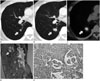
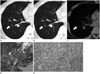
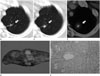
A 64-year-old man presented with incidentally detected pulmonary nodules on chest computed tomography (CT) images. He had a history of total thyroidectomy for thyroid cancer, 3 years prior, without recurrence. He was on medication for hypertension and asthma for 10 years. He was a non-smoker and did not have any pulmonary symptoms. Routine laboratory tests were within normal range. There were 3 lesions in both lungs on the CT images. Lesion 1 was a 0.7 cm solid nodule in the right lower lobe (RLL) superior segment (Fig. 1A). Lesion 2 was a 1.4 cm pure ground-glass opacity nodule located in the RLL lateral basal segment (Fig. 2A). Lesion 3 was an irregular-shaped 1.2 cm cavitary nodular lesion located in the left upper lobe (LUL) (Fig. 3A). There were no enlarged lymph nodes in the mediastinum.
At the 9-month follow-up, CT images showed enlargement of lesion 1 to 1.8 cm and the appearance of lobulation and spicule formation (Fig. 1B), whereas lesion 2 remained unchanged (Fig. 2B). The lesion 3 was also increased in size to 1.4 cm (Fig. 3B). Whole-body positron emission tomography-CT showed increased 18F-fluorodeoxyglucose (FDG) uptake in lesions 1 [standardized uptake value (SUV) = 7.4] and 3 (SUV = 3.8), but no FDG uptake in lesion 2 (Figs. 1C, 2C, 3C).
Pathological examination on biopsy specimens retrieved by CT-guided needle aspiration indicated that lesion 1 was non-small cell lung carcinoma. One week after biopsy, lobectomy of the RLL with mediastinal lymph node dissection, and wedge resection of the LUL of the lung were performed by video-assisted thoracic surgery. Macroscopic examination of the RLL of the lung revealed a 1.6 cm solid mass in the superior segment (Fig. 1D) and a 1.0 cm ill-defined lesion with discoloration in the lateral basal segment (Fig. 2D). Histopathologically, lesion 1 was confirmed as adenocarcinoma composed of solid (90%) and micropapillary (10%) patterns (Fig. 1E). Lesion 2 was adenocarcinoma, acinar predominant type (Fig. 2E). A 1.7 cm solid mass extending beyond the resection margin was found on cut sections of the LUL of the lung (Fig. 3D). Microscopic examination showed moderately differentiated squamous cell carcinoma (SCC) with visceral pleural involvement (Fig. 3E). Angiolymphatic invasion or lymph node metastasis was not detected.
There was no evidence of local recurrence on follow-up chest CT at 6 months post-chemotherapy.
DISCUSSION
The occurrence of synchronous triple primary lung cancers is very low and only a few cases have been reported previously (45). When detected simultaneously, only histologically different tumors are classified as multiple synchronous primary lung cancers; and in the case of the same histology, they should be considered as synchronous primary tumors based on features such as associated carcinoma in-situ or no evidence of lymphatic involvement or extrapulmonary metastases (1). During pre-operative staging, the importance of differentiating between multiple metastatic lesions and synchronous tumors is essential for tumor staging and appropriate treatment planning, but there is a lack of universal guidelines for diagnosing multiple synchronous lung cancers based on histopathological characteristics (56). However, the distinct radiologic features of multiple lung cancers could provide clues in the differential diagnosis of multiple synchronous versus metastatic lung cancers.
We reported the case of 3 synchronous tumors of the lung with different histology and radiologic features.
The lesion 1 was confirmed as a solid, predominant-type of adenocarcinoma. It appeared as a round solid nodule with a tumor doubling time of 203 days concomitant with lobulation and spicule formation, which are characteristic imaging findings of adenocarcinoma due to its desmoplastic reaction (7).
The lesion 2 was a persistent part-solid ground-glass nodule with a solid component in the central area, confirmed as acinar component predominant adenocarcinoma. On CT findings, part-solid nodular ground glass opacity suggests a malignancy rate of 64% including adenocarcinoma in-situ, minimally invasive adenocarcinoma or invasive adenocarcinoma (8).
The lesion 3 contained a cavity and had an irregular margin without calcification, compatible with SCC. The cavitary nodule with an irregular or lobulated border is one of the radiologic abnormalities of SCC (9). In SCC, approximately 10% show cavitation on CT because of central necrosis.
According to the Martini-Melamed criteria, these tumors are triple primary lung cancers owing to different morphologic patterns between lesions 1 and 2 (solid predominant vs. acinar predominant) and a different histologic subtype of SCC for lesion 3. There are some attempts to make up for the limitations in diagnosis of multiple synchronous lung cancers according to the criteria of Martini and Melamed (4). However, only a few reports deal with the imaging findings of each lesion in synchronous triple lung cancers (5). In our patient, the different microscopic findings for each of the synchronous triple primary lung cancers could be explained by their distinct radiologic characteristics, which correlated to the histologic subtypes reported in other studies (789). In addition to histopathologic criteria, distinguishing the different radiologic features of each lesion in synchronous primary lung cancer may be beneficial for diagnosis of the cancer.
 XML Download