

 PDF
PDF ePub
ePub Citation
 Print
Print


INTRODUCTION
Although it is important to diagnose the cardiac etiology in patients presenting with chest pain, non-cardiac causes of chest pain may also be serious. For many patients with chest pain who enter the emergency department or outpatient clinic, there is a non-cardiovascular cause of the pain (1). Indeed, among patients with chest pain who present for the first time to ambulatory care or the emergency room, only 11-39% are ultimately diagnosed with coronary artery disease (12).
A diagnosis of non-cardiovascular chest pain (NCCP) is made primarily from the patient's history, laboratory findings, and an imaging study. However, patient history and laboratory findings are of limited value for revealing the cause of pain. Thus, imaging findings are the most important factor in evaluating NCCP. The heart CT protocol is becoming the first-line evaluation tool for detecting significant coronary artery stenosis, with a high sensitivity of > 80%, and specificity > 90% (34). In contrast, assessment of NCCP remains a significant challenge, and conventional simple chest radiographs and routine CT are the first-line imaging tools.
Reported causes of NCCP include gastro-oesophageal, musculoskeletal, and pulmonary disorders, in addition to pleural illnesses and neurological and psychological problems. Tuberculosis (TB) is one of the diseases exhibiting these pulmonary disorders or pleural illnesses. Imaging findings for patients with NCCP as a presenting feature of TB varies, depending on the location of the lesion (pleura, subpleural lung, chest wall), activity of the lesion (active vs. stable), and the presence of effusion. NCCP associated with TB is relatively common in Korea, a region where TB is still endemic, but having a good prognosis (5). However, patients with NCCP often cannot work because of persistent pain and anxiety, interference in their daily activities due to pain, or because of the required repeated visits to the hospital (6). Although these patients undergo tests that exclude cardiac chest pain as a cause, considerable anxiety about the unrevealed cause of pain increases the health care visits and medical expenses. Thus, identifying the cause of the chest pain in patients complaining of persistent NCCP, would allow them to live more stable lives and spare them undue anxiety (7). Therefore, clinicians and radiologists should be more aware of typical CT findings, the frequency of NCCP, and the time interval from the initial TB diagnosis when evaluating the cause of NCCP, especially in regions where granulomatous disease is common. Few studies have examined chest CT findings of TB as the cause of NCCP, even though CT is an increasingly popular initial diagnostic tool for determining the cause of NCCP. Therefore, this study reviewed the chest CT findings in cases in which the causes of NCCP were confirmed, focusing on the clinicoradiological findings in cases of TB, in a region where the granulomatous disease is common.
MATERIALS AND METHODS
Patients
This retrospective study was approved by the Institutional Review Board. At our institute, chest pain is classified into four main categories: definitely, probably, probably not, and definitely not coronary chest pain. These clinical categories are based on the patient's chief complaint, symptoms and signs, risk factors, tests, and electrocardiogram findings. Cardiac CT is performed in patients with 'definite' or 'probable' cardiac chest pain at our hospital, and these patients were excluded from this study. Among the 409 patients who met the 'probably not' or 'definitely not' coronary chest pain criteria for enrolment, 211 (211/409, 51.6%) patients who had no significant CT findings and a final diagnosis of clinically insignificant chest pain, were also excluded. In addition, 36 cases with potentially life-threatening causes, such as aortic dissection and pulmonary embolism, for which a specific protocol was followed, or those with pericardial disease, were excluded. Finally, a total of 162 patients presenting to the outpatient clinic at our hospital, who had significant routine chest CT findings concordant with the final diagnosis for their NCCP, were included. Among the 162 patients who met all criteria for enrolment, 98 (60.5%) were male and 64 (39.5%) were female, with a mean age of 51 (range 18-88) years. All patients had significant CT findings, consistent with the final diagnosis. The chest CT study was conducted early in the diagnosis of these patients who had chest pain of unknown etiology, after simple radiography had been done.
Image Acquisition
Chest CT was performed using a 16-channel (n = 101) or 64-channel (n = 24) multi-detector CT (MDCT) scanner (Sensation; Siemens Medical Solutions, Erlangen, Germany). The parameters for 16-channel MDCT imaging were 120 kVp, 80-100 mAs, 3 mm thickness, and 1.5 mm collimation. Contrast-enhanced chest CT was obtained after injection of 30 g of iodinated contrast agent (100 mL iopromide, Ultravist 300; Bayer Healthcare Pharmaceuticals, Wayne, NJ, USA) at a rate of 2.3 mL/s using a power injector (OP100, Medrad, Pittsburgh, PA, USA). The parameters for 64-channel MDCT imaging were 120 kVp, 100 mAs, 3 mm thickness, and 1.2 mm collimation. Contrast-enhanced chest CT was obtained after injection of 30 g of iodinated contrast agent (100 mL iopromide, Ultravist 300) at a rate of 2.3 mL/s using a power injector (Stellant, Medrad). Scan data were displayed directly on two monitors (512 × 512 image matrix, 12-bit viewable grey scale) of a picture archiving and communication system (Starpacs, Infinitt, Seoul, Korea). A low-dose protocol (n = 43) was also performed using 16- and 64-channel MDCT scanners. The parameters were 100-120 kVp, 40 mAs, 3 mm thickness, and 1.2 mm collimation, without injection of a contrast agent.
Image Interpretation
Images were evaluated for causes of NCCP by location (rib, pleura, and subpleural lung) and the lesion type in each location. Specific non-cardiovascular lesion entities that were evaluated by location, included the following: rib lesions (fractures, malignant/benign tumors, and osteoarthritis), subpleural lung lesions (pneumonia, active/stable TB, atypical infection including parasite or fungus, and cancer), pleural lesions (TB/non-specific fibrotic pleural thickening, malignant pleural thickening, empyema, TB/bacterial or viral pleuritis, and pleuritis caused by connective tissue disorder), and pneumothorax. Fibrotic pleural thickening was defined as a continuous sheet of pleural thickening of greater than 2 mm thickness without effusion, and a definite adjacent parenchymal lesion with or without internal calcification. We defined the findings of "wet pleurisy" as pleuritis, which demonstrated focal or diffuse pleural thickening greater than 2 mm with effusion, on CT.
All imaging observations were decided by consensus of two radiologists. A final CT assessment for the cause of NCCP was recorded on a case report form.
In addition to the chest CT, information on other relevant diagnostic tests were also recorded, comprising sputum culture (including acid-fast bacilli), pleural fluid analysis, and any biopsy obtained within 1 month of presentation. A consensus group consisting of two radiologists and one physician from each clinical department was involved. Correlations were made between the presence of significant findings on CT and the final clinical diagnosis based on the history, examination, and any subsequent work-up up to the 1-month follow-up by the consensus group. We considered that the presumed CT lesions corresponded to the cause of the NCCP, when the pain improved after treatment of the targeted lesion.
RESULTS
Causes of Non-Cardiovascular Pain on Chest CT
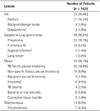
Locations and lesions causing NCCP are summarized in Table 1. The most common location for the cause of NCCP, as identified by CT, was the pleura (73/162, 45.1%), followed by subpleural lung parenchyma (49/162, 30.2%) and the ribs (33/162, 20.4%). The most common lesion that caused NCCP was TB (total, 54/162, 33.3%; stable TB, 40/162, 24.7%; active TB, 14/162, 8.6%) followed by pneumonia (31/162, 19.1%) and rib fracture (27/162, 16.7%). Other causes of NCCP were non-specific fibrotic pleural thickening (16/162, 9.9%), bacterial or viral pleuritis (9/162, 5.6%), and pneumothorax (7/162, 4.3%).
Among the NCCP cases as a result of rib involvement (33/162, 20.4%), sub-acute rib fractures were noted in 4 (2.5%) cases, and acute fractures were observed in 23 (14.2%) cases. A subpleural-located atypical infection as the cause of NCCP was demonstrated in 3 cases: 2 cases of fungal pneumonia, and 1 of Paragonimus Westermani infection.
Tuberculosis
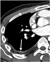
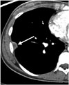
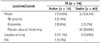
Of the 54 TB cases, 40 (74.1%) were stable TB and 14 (25.9%) were active TB. Location of TB as the cause of NCCP is summarized in Table 2. NCCP in TB patients was most commonly located in the pleura (39/54, 72.2%). The most common lesions encountered were fibrotic pleural thickening (30/54, 55.6%) (Fig. 1), acute and chronic empyema (5/54, 9.3%) (Fig. 2), and TB pleuritis (4/54, 7.4%). Subpleural lung lesion (14/54, 25.9%) was the second most common location of NCCP, in both stable pulmonary TB (8/54, 14.8%) and active pulmonary TB (6/54, 11.1%). A rib lesion was the cause of NCCP in 1 case of TB (1.9%). Thus, the most common condition causing NCCP in patients with TB was fibrotic pleural thickening in stable TB, followed by subpleural stable pulmonary TB.
Active TB
For patients with active TB, pleural lesions were noted in 7 (13.0%) cases, subpleural lung lesion in 6 (11.1%) cases, and there was 1 case (1.9%) of rib lesion. Among the pleural lesions, TB pleuritis was diagnosed in 4 cases and localized TB empyema was present in 3 cases (Fig. 2). A small amount of pleural effusion, which was not detected on simple radiographs, was seen in all cases with TB pleuritis. For subpleural lung lesions, typical conglomerated nodules with a tree-in-bud appearance adjacent to the pleura were noted in 5 cases, and 1 case had patchy necrotic consolidation, mimicking pneumonia. The rib lesion involved the right sternocostal junction and was not evident on simple radiographs.
Stable TB
In stable TB patients, pleural lesions were noted in 32 (59.3%) cases and subpleural lung lesions in 8 (14.8%) cases. There was no case involving a rib lesion as the cause of NCCP. All pleural lesions causing NCCP in stable TB showed fibrotic pleural thickening, and calcification was also seen in 21 cases. All subpleural lung lesions also showed typical stable TB features, with fibrotic bands adjacent to the pleura.
Among the TB cases, 18 had a known duration from the day of initial TB diagnosis to the day that chest pain developed. The mean duration was 2.1 months (range, 15-360 days) for active TB (n = 12) and 46.8 months (range, 31-84 months) for stable TB (n = 6). In active TB cases, the mean duration was 1.9 months for pleural lesions (n = 7) and 2.5 months for subpleural lung lesions (n = 5).
DISCUSSION
In this study, lesions that caused NCCP were investigated, mainly in cases of acute/chronic pulmonary or pleural infections, with the exclusion of so-called 'triple rule out' lesions. Major pathologies that produce NCCP include gastro-oesophageal reflux and potentially life-threatening causes, such as aortic dissection, pulmonary embolism, tension pneumothorax, and esophageal perforation (8). However, for diagnosis of these lesions as causes of NCCP, routine chest CT is either not indicated as the tool of choice (for gastro-oesophageal reflux, oesophageal perforation, and pneumothorax), or another CT protocol is performed (CT angiography for aortic dissection and thromboembolism). Thus, NCCP cases due to gastro-oesophageal reflux and potentially life-threatening cases were not included in this study.
Most cases of NCCP involve benign infectious/inflammatory conditions. A careful evaluation of the patient is necessary to diagnose the underlying disorder or lesion, since such benign causes tend to recur, especially in cases related to chronic lesions, and may lead to patient anxiety and higher costs.
The CT manifestations of NCCP cases caused by rib lesions were not surprising, as this is a well-known cause.
Our group was particularly interested in TB, which represented more than 30% of the causes of NCCP cases in this study. According to our study, in addition to the pleural changes and rib lesions from TB, the active stage of a subpleural pulmonary lesion was the cause of NCCP in 3.6% of the patients enrolled. As the patterns of this disease can mimic cardiac pain, these findings should be considered, particularly in areas with endemic granulomatous disease. Moreover, based on the high incidence of TB as the cause of NCCP in our study, we suggest that the prevalence of NCCP might increase in areas where the granulomatous disease is endemic.
Previous investigations that have evaluated chest pain related to pleural thickening are limited to those concerning benign asbestos pleural disease or mesothelioma, rather than pleural tuberculous lesions (89). Yates et al. (10) has reported on the incidence of chest pain (56%) with diffuse pleural thickening related to asbestos.
We observed that in TB patients, the most painful location was over the thickened pleura. This might be caused by irritation of the sensory nerves of the parietal pleura or the intercostal nerves, since they are not protected by the internal intercostal muscles. Kim et al. (11) reported CT findings of thickened pleura in patients with chronic TB empyema and correlated their findings with those from histopathology. Their study determined that, within the pleura, deeper mesothelial and submesothelial fibroelastic layers were thickened, with fibrin deposition and ingrowth of capillaries and fibroblasts (12). We suggest that these histopathological changes in stable TB correlate with pleural thickening; the fibrotic band and lung parenchymal distortion, observed on CT, would be more likely to irritate the sensory nerves of the parietal pleura and increase the frequency of NCCP in stable TB, as compared with active TB. We believe that further studies, involving a larger study group, are needed to investigate the incidence and precise mechanisms underlying the chest pain due to pleural thickening caused by TB.
In addition to a higher proportion of pleural thickening, we found that the subpleural lung parenchymal lesions due to active TB were also associated with NCCP. However, we are unable to explain the exact pathogenesis of these lesions as a cause of pain. Nevertheless, because the location of the NCCP was consistent with the subpleural lung lesions detected by CT and the pain disappeared after the lesions improved, we believe that these lesions were the cause of the chest pain. We suggest that the organizing stage of pulmonary TB or pneumonia, with ingrowth of fibroblasts along the fibrin sheets lining the adjacent parietal pleura, might be a cause of pain.
Pleuritic pneumonia has been reported as a major cause of NCCP, with typical bacterial, viral, or fungal infections. NCCP has been reported as a less common major symptom in patients with "bronchiolitis obliterans organizing pneumonia" (BOOP). In particular, in approximately 50% of patients with radiological manifestation of the multiple nodular type of BOOP, the mode of presentation was NCCP (13). In our study, 2 patients with pneumonia had confirmed BOOP, and both showed the nodular pattern on CT. There were no cases of chest CT-positive mediastinal or pericardial disease as a main cause of NCCP in this study.
One limitation of this study was that the decision regarding the etiology of the NCCP was made subjectively by an agreement among two radiologists and one clinician, raising the need for a study with more observers. However, we believe that the results of this study are reliable, because the pain resolved after the targeted lesion was treated. The possibility of radiating pain from another cause could result in an erroneous decision regarding the cause of NCCP, as a coincidental lung or pleural lesion cannot be excluded. As we included only CT-positive subjects, many patients with gastro-oesophageal reflux, which is one of the main causes of NCCP and a life-threatening lesion, were excluded from this study. Additionally, intra-abdominal causes were also excluded. Therefore, the incidence of each of the causes of NCCP examined in this study might not represent the true percentage distribution among all causes of NCCP.
In conclusion, the chest CT evaluation of patients revealed that stable TB and subpleural pneumonia were the major causes of NCCP. Focal fibrotic pleural thickening from stable TB was the most common cause (23.8%) of NCCP detected with chest CT, taking into consideration benign infectious/inflammatory causes of NCCP in a TB endemic area.
 XML Download