

 PDF
PDF ePub
ePub Citation
 Print
Print


INTRODUCTION
Chaussy et al. (1) first described extracorporeal shock wave lithotripsy (ESWL) in 1980 and it has since become a well-established treatment for urolithiasis. Approximately 3-7% of patients have complications after ESWL, which are usually mild, however more severe conditions, such as life-threatening kidney injury or retroperitoneal hemorrhage, is extremely rare (2). To the best of our knowledge, in this study we report the first case of a patient with emphysematous pyelonephritis (EPN) after ESWL, who expired after a complicated clinical course.
CASE REPORT
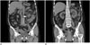
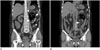
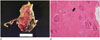
A 65-year-old woman visited the hospital because of the left flank pain. The patient had a past medical history of diabetes as an underlying disease. On the urinary analysis performed in different institution, white blood cell (WBC) was 10-20/high power field (HPF) and red blood cell was 5-10/HPF. The patient did not present symptoms of a urinary tract infection. A non-enhanced computed tomography (CT) scan showed a 7-mm sized stone in the left proximal ureter with diffuse swelling of left kidney, hydronephrosis, and perinephric infiltration (Fig. 1). Acute pyelonephritis cannot be ruled out without enhanced CT scan, but there was no evidence of EPN, such as destructed renal parenchyma. The patient underwent an ESWL for proximal ureter stone. The ESWL was performed using a standard treatment protocol: 3000 shock waves with a total energy of 180 J/cm2. Abdominal pain and oliguria developed soon after the procedure. The patient was transferred to the Emergency Department with persistent abdominal pain despite taking analgesics. Her blood pressure was 128/75 mm Hg and body temperature was 37.7℃. On laboratory analysis, the WBC was 19.08 × 10⁹/L, the platelet count was 15 × 109/L, the C-reactive protein was 331.52 mg/L, the erythrocyte sedimentation rate was 36 mm/h, and creatinine, 5.46 mg/dL. Urine analysis was not performed because of anuria. A non-enhanced CT scan of the abdomen showed gas replacing the left kidney parenchyma, which was ultimately destroyed, and extension of gas to perinephric space. A left proximal ureteral stone was still noted without any signs of breakage (Fig. 2). An emergency left nephrectomy was performed after the diagnosis of EPN was made. After surgery, the patient was anuric and did not maintain blood pressure. Then patient underwent continuous renal replacement therapy, but did not maintain blood pressure and developed sepsis. The following day the patient expired. E. coli were reported in the blood culture and diffuse necrosis, hemorrhage, and focal abscess formation in the renal parenchyma, which was consistent with renal infarction, were reported in the pathology report (Fig. 3).
DISCUSSION
Complications of ESWL are related mostly to residual stone fragments, infections, and effects on tissues, such as urinary, gastrointestinal, cardiovascular, genital, and reproductive systems (3). EPN, caused by ESWL, has not been reported in the literature, and furthermore the mechanism is still unknown. To the best of our knowledge this is the first case reported in the literature. The tearing and shear forces and cavitation activity produced by ESWL in tissue and vessels may cause the initial renal injury (3, 4), while expansion or collapse of bubbles in the vessels results in vessel damage (5, 6). There are four factors pertinent to the pathogenesis of EPN: the presence of a gas-forming organism, high serum glucose levels, impaired tissue perfusion, and compromised immune response. Diabetic patients are predisposed to these factors, which may be worsened in the setting of ureteric obstruction (7, 8). Thus, the mechanism of EPN after ESWL, in a patient with diabetes mellitus, can be hypothesized as being caused by renal parenchymal and vessel injury with a superimposed gas forming a bacterial infection. In this case, the pyuria was not initially deemed significant because the patient did not present symptoms of urinary tract infection, which is an absolute contraindication for ESWL.
EPN does not have characteristic symptoms and signs, thus, radiologic findings are crucial factors in distinguishing EPN from the less serious cases of pyelonephritis. There are two types of EPN. Type 1 EPN is characterized by renal parenchymal destruction that manifests with either streaky or mottled areas of gas but no fluid collections. Type 2 EPN is characterized by renal or perirenal fluid collections that are directly associated with bubbly or loculated gas or by gas within the urinary collecting system. The mortality rate for type I EPN is higher (69%) than that for type II (18%) (9). Prognosis of EPN is generally variable. Type I EPN, bilateral EPN, thrombocytopenia, and conservative treatment are associated with increased mortality in patients with EPN. In addition, increased serum creatinine level, disturbance of consciousness, and hypotension (systolic blood pressure less than 90 mm Hg) may also be risk factors (10). In this case, the patient expired earlier than expected, most likely because of the presence of type I EPN, thrombocytopenia, increased serum creatinine level, disturbance of consciousness, and hypotension. Emergent surgical drainage or even nephrectomy may be necessary for severe cases. If conservative treatment is not effective, surgical treatment is required as in this case. The most important factor is early diagnosis. An abdominal CT scan is the preferred diagnostic tool because it can measure the extent and amount of gas and degree of renal parenchyma destruction. In patients with suspected immediate inspection should be performed, including urgent CT.
When diabetic patients present with abdominal pain and fever after the development of ESWL, physicians should consider the possibility of EPN and should closely observe the patient for clinical signs that are suggestive of septic shock.
 XML Download