

 PDF
PDF ePub
ePub Citation
 Print
Print


INTRODUCTION
Bronchial artery embolization (BAE) is an effective and widely used treatment in patients with massive, recurrent hemoptysis, particularly when it is related to inflammatory lung diseases such as acute and chronic pulmonary tuberculosis, bronchiectasis, and aspergillomas (1).
Anomalous bronchial arteries occur frequently at ectopic sites other than the upper descending thoracic aorta. There are various aberrant bronchial arteries, and precise anatomical knowledge of these variations is necessary prior to BAE. For an accurate evaluation of massive hemoptysis, computed tomography (CT) is usually employed and is helpful in localizing the site of bleeding and in detecting the bronchial arteries, including those with anatomical variations. Moreover, CT is valuable for determining the underlying disorder inducing the hemoptysis (23). Recently, multidetector computed tomography with angiography is being used more often. The anatomy of the bronchial artery is often beautifully demonstrated with these combined techniques, which can be very helpful prior to angiography (45). We encountered an extremely rare case of aberrant left bronchial artery arising from a replaced left hepatic artery in a 70-year-old man with massive hemoptysis.
CASE REPORT
A 70-year-old man was admitted to the emergency department with hemoptysis for the past 3 days. After he arrived at our hospital, he experienced three episodes of hemoptysis, totaling approximately 300 mL. He had a 3-year history of bronchiectasis and reported having had a frequent cough for several months that was productive of yellowish, sometimes blood-tinged sputum.
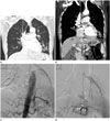
Contrast-enhanced chest CT revealed bronchiectasis in both lobes, but predominantly in the left lower lobe (Fig. 1A). Axial CT images showed two dilated bronchial arteries, each traveling to respective right and left pulmonary hila; however, the origin of the left bronchial artery could not be found in its normal anatomical position. A coronal reformatted image revealed a tortuous artery branching from a replaced left hepatic artery and traveling upward along the descending thoracic aorta (Fig. 1B).
The right common femoral artery was accessed percutaneously, and upper thoracic aortography with a 5-Fr pigtail catheter (Cook, Bloomington, IN, USA) was carried out. The right intercostobronchial trunk and the right bronchial artery were identified, and the two right bronchial arteries were catheterized. Although the left bronchial artery did not appear to be in its normal position relative to the thoracic aorta, a tortuous vessel with an ascending course was faintly visible on repeat lower thoracic aortography (Fig. 1C). Therefore, we suspected that it had an anomalous origin, so we performed selective angiography of the branches arising from the upper abdominal aorta. Celiac angiography with a 5-Fr Rosch hepatic catheter (Cook) showed a replaced left hepatic artery arising from the left gastric artery and the left bronchial artery arising from the replaced left hepatic artery (Fig. 1D). A hypertrophied and tortuous left inferior phrenic artery was also seen on the selective angiogram.
Selective embolization was performed using a 2.2-Fr microcatheter (Progreat, Terumo, Tokyo, Japan) to introduce polyvinyl alcohol particles (Contour, Boston Scientific, Natick, MA, USA) with a diameter of 350-500 µm via the right intercostobronchial trunk, the right bronchial artery, the replaced left bronchial artery, and the left inferior phrenic artery.
The patient received conservative and antibiotic treatment for bronchopneumonia with underlying bronchiectasis. Six days after the procedure, he continued to have an intermittent cough with a small amount of hemoptysis, and the conservative therapy was continued. He was discharged about 2 weeks later without complications.
DISCUSSION
Massive hemoptysis is an emergent condition, and the physician must decide whether interventional management is needed (3). In most cases, massive hemoptysis originates from the bronchial rather than the pulmonary circulation, so BAE is the preferred management. However, non-bronchial systemic arteries may also induce hemoptysis (5).
BAE was first introduced by Remy et al. (6) in 1973 and has since been used to manage massive and recurrent hemoptysis. The bronchial artery usually originates from the descending thoracic aorta between the lower margin of T4 and the upper margin of T6 (7). Aberrant or anomalous bronchial arteries are defined as those originating at any other level. Cauldwell et al. (7) reported in 1948 that in their examination of 150 human cadavers, 16.7% of the bronchial arteries originated from ectopic areas. Because aberrant bronchial arteries extend along the major bronchi, they can be distinguished from non-bronchial systemic collateral vessels. The most common aberrant bronchial arteries arise from the aortic arch (789). Anomalous bronchial arteries may originate from the subclavian, internal mammary, brachiocephalic, vertebral, carotid, pericardiacophrenic, coronary, esophageal, left gastric, and inferior phrenic arteries; the thyrocervical and costocervical trunks; and the lower descending and abdominal aorta (8910). A bronchial artery arising from a replaced left hepatic artery has not been reported, while two cases have been reported of bronchial arteries arising from the left gastric artery (810). However, Ponnuswamy et al. (4) described a case of additional vessels arising from non-bronchial systemic collateral arteries stemming from the celiac trunk and hepatic artery (4).
In our patient, aortic arch angiography did not show the left bronchial artery in the usual position. At celiac angiography, a certain artery seen on CT arose from a replaced left hepatic artery and coursed over the lower margin of the left bronchus at the level of the carina. Therefore, this is not an artery originating from non-bronchial collateral vessels, but an aberrant bronchial artery originating from an unusual site. Non-bronchial systemic arteries enter the lung parenchyma through the inferior pulmonary ligament or the adherent pleura, and their course does not parallel that of the bronchi.
Jiang et al. (8) and In et al. (10) reported two cases of replaced left bronchial arteries arising from the left gastric artery. According to these authors, the cause of this variation was occlusion of the left bronchial artery due to extensive disease of the left lung, and it was then reconstituted via collateral flow of the esophageal artery from the left gastric artery. Similarly, with respect to our patient, we assume that the left bronchial artery might be reconstituted by collaterals from the replaced left hepatic artery that had arisen from the left gastric artery.
In conclusion, when the bronchial artery of the affected side of the lung is not found on angiography for BAE despite all possible efforts, one must suspect an anomalous origin, and the hepatic artery should be considered a possible site.
 XML Download