

 PDF
PDF ePub
ePub Citation
 Print
Print


INTRODUCTION
Spontaneous breast infarction is a very rare episode, representing about 0.1% of biopsied (1) and 0.3-1.5% of fibroadenoma cases including the reported cases of pregnant women (234). Although several cases of breast infarction have been reported in the literature, we found only two reports that described the mammographic findings of an infarcted breast tumor involving five patients (15). In the present study, we describe a case of spontaneous breast infarction in a postpartum woman and discuss the mammography and ultrasonography imaging findings.
CASE REPORT
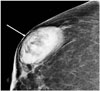
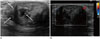
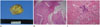
A 33-year-old woman with a palpable mass on her right breast visited our hospital four weeks postpartum. She initially discovered the palpable breast mass during her 3rd trimester of pregnancy and reported no change in its size after delivery. She did not experience nipple discharge or pain and had no history of trauma or use of anticoagulant drugs. The physical examination revealed a hard and non-tender mass in the right breast, without nipple retraction and skin thickening. The patient had no axillary or supraclavicular lymphadenopathy. Mammography revealed a large and oval shaped fat-containing mass with a circumscribed margin and microcalcifications (Fig. 1). On ultrasonography, the mass was seen as an oval shaped structure with a circumscribed margin and heterogeneous echogenicity along with a combined pattern of posterior features (Fig. 2A). A few echogenic dots were observed within the mass. Minimal vascularity was noted at the periphery of the mass on a color Doppler image (Fig. 2B). Considering these radiologic findings as probably benign, the mass was classified as a category 3 lesion. However, the patient wanted to perform biopsy for the fear of malignancy. The mass was pathologically verified as a breast infarction. Three months later, there was no change in size of the mass and the patient underwent excisional biopsy. The specimen was reported as a circumscribed solid mass measuring 4.8 × 3.6 × 2.3 cm with necrosis on the cut surface (Fig. 3A). There was extensive necrosis with adjacent fibrosis and residual epithelium, as well as lactation hyperplasia (Fig. 3B). Microcalcifications and cystic changes were also seen within the mass (Fig. 3C). There was no evidence of malignancy and the histologic examination confirmed the features of infarcted hyperplastic breast tissue.
DISCUSSION
Spontaneous breast infarction is a very rare complication of fibroadenoma of the breast. According to a recent study by Oh et al. (1), 0.1% of all biopsied lesions (9/8310) were spontaneous infarctions. Infarctions in benign lesions may occur in various conditions such as fibroadenoma, intraductal papilloma, and hyperplasia associated with pregnancy and lactation, as well as during anticoagulant therapy and with oral contraceptive use (123678). The malignant etiologies of breast infarction include invasive carcinoma, ductal carcinoma in situ, as well as medullary carcinoma, and papillary carcinoma (19). Spontaneous breast infarction may occur as an uncommon complication of fine needle aspiration biopsy (10). The vascular trauma during the needling procedure may induce thrombosis and infarction.
The presenting symptom is most frequently a rapidly growing palpable mass that is sometimes painful and hard. Occasionally, bloody discharge secondary to tissue necrosis can be observed (7). In such patients, the clinical diagnosis of an inflammatory or malignant breast disease may be possible. The cytopathologic features of infarction include necrosis and worrisome nuclear features, which are often confused with inflammation or malignancy (8).
Spontaneous infarction has been mostly described during the third trimester of pregnancy and lactation. In these cases, the proposed pathogenesis includes a relative vascular ischemia during phases of increased metabolic requirements of the breast tissue (3). The possibility of a probable torsion due to the increased mobility of these tumors has been suggested in cases of spontaneous infarction occurring in the absence of pregnancy and lactation.
Until date, we were able to locate only two reports that described the mammographic findings of an infarcted breast tumor involving five patients. In one study, an infarcted lactating adenoma presented as a circumscribed mass without additional findings on mammography (5). Two infarcted fibroadenomas and one medullary carcinoma similarly exhibited oval shaped, indistinct, and highly dense masses (1). An infarcted intraductal papilloma presented as a round, circumscribed, equal density mass on mammography. As indicated by these reports, it is evident that no characteristic mammographic findings lead to differentiation between malignant and benign tumor infarctions of the breast.
There are a few reports on the ultrasonographic findings of spontaneous breast infarction. On ultrasonography, the most common reported features include an oval shaped, parallel-oriented, indistinct, heterogeneously hypoechoic mass with or without posterior acoustic enhancement. In addition, these lesions are sometimes irregular shaped and angularly margined mass that can be shown for malignancy.
In conclusion, we report a recent case of infarcted hyperplastic breast tissue in a young postpartum woman. The spontaneous breast infarction presented as a circumscribed mass with microcalcifications on mammography, and a heterogeneous echoic mass with echogenic dots on ultrasonography. There was minimal vascularity observed at periphery of the mass on color Doppler imaging. It is proposed that even though the incidence of a spontaneous breast infarction is very low and it has no specific radiologic features, it should be included in the differential diagnoses of women with a recent history of pregnancy or lactation.
 XML Download