

 PDF
PDF ePub
ePub Citation
 Print
Print


INTRODUCTION
A fibro-osseous pseudotumor is an extremely rare benign ossifying subcutaneous mass, which usually presents as an ill-defined soft tissue mass with focal calcification, particularly in the proximal phalanx of the digit. It usually occurs in young adults, with a slight predominance in females, and presents as a painful and localized soft tissue swelling of the finger (12). This lesion has been considered to be the subcutaneous counterpart of myositis ossificans, and the clinical and histological features thereof can be confused with those of a malignant tumor, particularly an extraskeletal osteosarcoma. Accurate interpretation of clinical, radiological, and histological features is very important to avoid misdiagnosis and unnecessary amputation (34). A fibro-osseous tumor is benign, and has an excellent prognosis without local reccurence, after complete excision (45).
To date, only a few case reports have described the magnetic resonance imaging (MRI) findings of this rare lesion (67), thus less is known about the MRI features of fibro-osseous pseudotumor.
Therefore, here, we describe the radiological features, including MRI findings, of a case of a fibro-osseous pseudotumor that involved the subcutaneous tissue of the proximal phalanx of the digit, in a 19-year-old female.
CASE REPORT
A 19-year-old female presented with a rapidly growing, palpable painful mass on the digit of the right hand that had commenced 12 months prior. She had no history of trauma in that area and was otherwise healthy.
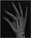
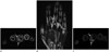
On physical examination, a firm and non-movable mass could be palpated on the radial side of the proximal phalanx of the right middle finger. Laboratory data, including the erythrocyte sedimentation rate and C-reactive protein level, were unremarkable. There was no erythema, ulceration, or infection of the overlying skin. Radiography revealed a well-circumscribed, ossified soft tissue mass, adjacent to, but separate from the bone, overlying the proximal phalanx of the right middle finger, but without any periosteal reaction or bony erosion (Fig. 1). To evaluate the relationship of the mass to adjacent soft tissues and bone, and to characterize and assess the extent of the mass, MRI of the right hand was performed using a 3.0-T scanner (Magnetom Skyra, Siemens, Erlangen, Germany). A well-defined, elongated ovoid mass was observed overlying the proximal phalanx of the right middle finger, measuring approximately 17 × 10 × 16 mm. It was clearly separate from the nearby proximal phalangeal bone, growing principally into subcutaneous tissues (Fig. 2).
On T1-weighted and T2-weighted images, the mass displayed high signal intensity relative to that of skeletal muscle, with areas of intermediate signal intensity. No periosteal reaction or cortical change in adjacent bone was evident (Fig. 2A, B). At first, the hyperintense area of the mass was thought to be a fatty change or hemorrhage. After contrast enhancement, the hyperintense area on T1- and T2-weighted images became hypointense and exhibited little enhancement on fat-suppressed contrast-enhanced T1-weighted images, suggesting a fatty change. However, the intermediate-signal area on T1- and T2-weighted images exhibited high contrast enhancement (Fig. 2C). As the mass contained a fatty component, several calcifications, and foci of contrast enhancement, our initial diagnostic impression was a soft tissue hemangioma.
During surgery, we removed a 15 × 10 × 15-mm calcified mass that was clearly separate from the phalangeal bone and the flexor sheath. The lesions were well-circumscribed, grayish-white, and firm. Surgical excision was complete. There was no evidence of tenosynovitis or adhesion.
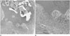
On gross examination, the resected specimen was found to be an irregularly lobulated bony tumor in subcutaneous fat tissue. Microscopic examination showed that the lesion had a peripheral fasciitis-like stroma of fibroblasts and a central irregular immature osteoid. There were mild to moderate cellular proliferation of fibroblasts at the periphery and immature osteoid formation with osteoblastic rimming (Fig. 3).
The final diagnosis was a fibro-osseous pseudotumor of the digit. No further treatment was required. No recurrence was noted during the 12-month follow-up.
DISCUSSION
A fibro-osseous pseudotumor of the digit is a rare benign lesion exhibiting reactive fibroblastic proliferation with foci of osseous differentiation. The lesion was first described by Dupree and Enzinger (8) in 1986 as a reactive rather than a neoplastic lesion. Clinically, the lesion primarily affects young adults, with a slight predominance in females, and presents as a rapidly growing, painful localized mass. The lesion originates in subcutaneous tissues and adjacent fibrous structures of the fingers, particularly in soft tissue of the proximal phalanx, and less commonly, the toe, without muscular involvement. The pathogenesis of fibro-osseous pseudotumors is not fully understood, however, repeated trauma seems to be recognized as a predisposing cause (58). Nevertheless, in our case, there was no history of trauma.
The clinical diagnosis of a fibro-osseous pseudotumor is difficult because of its rarity. The lesion can be confused with a malignancy because of its aggressive nature and rapid growth, resulting in unnecessary amputations (8). However, the lesion is a noninvasive entity and can be cured by simple resection.
The essential histopathological features of a fibro-osseous pseudotumor are localization in subcutaneous tissue without muscular involvement; an irregular multi-nodular growth pattern; the presence of a mixture of fibroblasts, osteoblasts, and osseous trabeculae with varying degrees of maturation; and the absence of the typical zoning pattern of myositis ossificans (78). A typical zoning pattern is the presence of immature cellular areas in the centre of the lesion, and more mature ossifying areas at the periphery (9).
Additionally, a fibro-osseous pseudotumor demonstrates focal hypercellularity, cellular atypia, and increased mitotic activity, which may trigger a mistaken pathological diagnosis of sarcoma (58). Plain radiographs revealed an ill-defined and calcified soft tissue mass in the digit.
Generally, the lesion was located close to, but clearly separate from, the underlying bone; a periosteal reaction is absent; and the musculature is not involved. These findings are very helpful to differentiate the tumor from other bone or soft tissue tumors (6810).
MRI may be helpful to evaluate the relationship of such a lesion with the underlying bone and soft tissue. However, less is known about the MRI features of a fibro-osseous pseudotumor than those of plain radiographs and histopathology.
In our case, MRI revealed a subcutaneous, well-circumscribed soft tissue mass adjacent to, but separate from, the bone. Retrospectively correlating the MR imaging with the pathological findings of our case, the digital soft tissue mass was heterogeneous in terms of signal intensity (intermediate and high) on T1- and T2-weighted images. The regions of intermediate signal intensity probably include the fibroblastic proliferative portion, whereas the hyperintense region is thought to be fatty marrow of the osseous portion (Fig. 2A, B).
On fat-suppressed contrast-enhanced T1-weighted imaging, the intermediate signal areas on T1- and T2-weighted images exhibited contrast enhancement, suggesting a fibroblastic proliferative area. On the contrary, the area of little contrast enhancement was thought to be the osseous portion (Fig. 2C). Our MRI findings were in good agreement with the pathological features of a fibro-osseous pseudotumor, which consists of mixed osseous and fibroblastic proliferation. In general, fibroblastic proliferative components exhibit low-to-intermediate signal intensities on T1-weighted images, variable signal intensities on T2-weighted images, and contrast enhancement on fat-suppressed contrast-enhanced T1-weighted images, whereas osseous components are hyperintense on T1- and T2-weighted images because of the fatty marrow, and show little contrast enhancement on contrast-enhanced T1-weighted images (6710).
The main differential diagnoses for a fibro-osseous pseudotumor include myositis ossificans, Turret exostosis, bizarre parosteal osteochondromatous proliferation (BPOP) of the bone (the Nora lesion), and extraskeletal osteosarcoma (15). A fibro-osseous pseudotumor shares histological and clinical features with myositis ossificans, and some authors regard the lesion as a subcutaneous variant of myositis ossificans (1810). A fibro-osseous pseudotumor occurs mainly in the subcutaneous tissue of a digit without muscular involvement, however myositis ossificans usually occurs in deeper soft tissues, and histopathologically exhibits a typical zoning phenomenon. Therefore, a distal superficial lesion lacking a zoning pattern will favor diagnosis of a fibro-osseous pseudotumor, while a proximal and deep lesion with a zonation pattern will suggest a diagnosis of myositis ossificans (4589).
Extraskeletal osteosarcoma should always be ruled out; this usually develops in a large muscle of the proximal lower extremity and affects patients of older age. Fibro-osseous pseudotumors are more common in young adults, and are located more distally.
Nevertheless, a fibro-osseous pseudotumor can be misdiagnosed as a malignant neoplasm, both radiographically and histopathologically. Both BPOP and Turret exostosis are similar to fibro-osseous pseudotumors in terms of clinical, histological, and radiological findings, being distinguished from fibro-osseous pseudotumors by attachment to underlying bone (by stalks) and endochondral ossification. A fibro-osseous pseudotumor is an extraosseous lesion (56810).
Morphologically, BPOP has a cartilaginous cap that may exhibit bizarre atypia, whereas cartilage is rarely evident in a fibro-osseous pseudotumor, which develops via intramembranous ossification without any cartilage intermediate. Both lesions can be preceded by trauma, but BPOP is usually painless, and a fibro-osseous pseudotumor painful (5).
Despite its benign nature, the recommended treatment for a fibro-osseous pseudotumor is complete surgical excision; the prognosis is excellent with no evidence of malignant transformation (12458).
The most important problem associated with a fibro-osseous pseudotumor is that it can mimic a malignancy because of rapid growth, hypercellularity, cellular atypia, and mitotic activity. Therefore, it is very important to clearly distinguish the lesion from a malignant neoplasm, particularly a sarcoma (7).
In summary, a fibro-osseous pseudotumor is a rare and reactive lesion of fibroblastic proliferation with focal bone formation, and usually occurs in the dermis and subcutaneous tissue of the digit, without muscular or bony involvement. Imaging findings are helpful in differential diagnosis, avoiding unnecessary treatment such as radical amputation.
 XML Download