

 PDF
PDF ePub
ePub Citation
 Print
Print


INTRODUCTION
Percutaneous abscess drainage has become a widely accepted procedure for the treatment of various abdominal or pelvic abscesses (1234). According to several studies, percutaneous abscess drainage is curative in 80-90% of cases (5678910). While the majority of cases are manageable, having a straightforward access, some cases may be inaccessible or undrainable due to limited topographic factors caused by anatomic distortions secondary to surgery, surrounding organs, and difficult location. To overcome an access limitation, invasive transgressions of surrounding organs may often be necessary. Occasionally, a surgical drain placed near the abscess during surgery, can be used as an alternative access route (11). However, even with alternative access route, some abscesses remain inaccessible and eventually require surgical drainage.
The recently developed C-arm cone-beam CT (CBCT) system, that enables imaging with wide z-axis coverage in 1 axial scan, offers a greater flexibility in orientating the detector around the patient, than the closed CT gantry systems (12). CBCT is space and time-saving because it is integrated with the C-arm gantry, which enables real-time CT during procedures without patient transfer to CT room. For these lesions, CBCT has been applied in various interventional procedures and surgeries for hepatic tumor embolization, guidance for biopsy, and surgeries for spine and skull base (131415161718).
Until now, there have been few studies on the use of CBCT in drainage procedure, and no reports with respect to drainage of inaccessible or undrainable abscess. CBCT is expected to allow the identification of the trajectory of needle and guide wire during procedures, which can improve the accuracy and efficiency of the process. In addition, real-time fluoroscopic capability of CBCT can reduce the procedure time, as compared with conventional CT fluoroscopic system because on-site imaging during interventional procedure is possible with CBCT. Thus, the purpose of this study was to evaluate the clinical usefulness of CBCT in drainage of inaccessible abscess.
MATERIALS AND METHODS
This retrospective study was approved by our Institutional Review Board and requirement for informed consent was waived.
Patient Characteristics
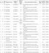
From September 2009 to February 2015, a total of 29 CBCTs were performed for 21 inaccessible abscesses (21 patients: 8 male and 13 female; mean age: 59 years) (Table 1).
CBCT Equipment and Parameters
CBCT was performed using a flat-panel detector C-arm system (XperCT, Allura FD20; Philips Heathcare, Best, the Netherlands). Two CBCT protocols (Abdomen Fast LD or Abdomen Roll) were followed. Three hundred and twelve images were acquired during 180-240° rotation of C-arm in a session of CBCT. Rotation time of C-arm was 5.2-10.4 seconds, depending on the scanning protocol used. Raw data was transferred to an external workstation and the images were reconstructed with 1.98 mm slice thickness.
Drainage Procedure with CBCT
The planning of each drainage procedure was performed by the attending interventional radiologist. Before each drainage procedure, the diagnostic CT scans were reviewed and the access route was determined, including skin puncture site and the needle path. The placement of surgical drains at the time of surgery, either near the abscess or fistula opening on skin, was chosen as an alternative access route.
Initially, local anesthesia (2% lidocaine hydrochloride) was administered at the skin puncture site and the planned needle path, using a 21-gauge needle. Local anesthesia was not administered when the surgical drain or fistula opening was used as an access route. During local anesthesia, skin puncture, and needle insertion, ultrasonography (US) was used for determination of the puncture site although the abscess was not clearly depicted on the US.
CBCT was indicated when 1) the safe access window to the abscess was narrow and there was a risk of transgression of adjacent or overlying organs, 2) the abscess was deeply seated and had a poor sonographic visualization, 3) the abscess had an irregular dispersed shape with a remote main segment, which made it difficult to localize the needle or guide wire to the main portion of the abscess, 4) the guide wire could not be inserted in the abscess, despite repeated trials.
In case of direct puncture, CBCT was performed after a 22-gauge Chiba needle was advanced to the target depth along the planned path, previously estimated by a diagnostic CT scan. When the surgical drain or fistula opening was used as an access route, CBCT was performed if the guide wire positioning to the main portion of the abscess failed, despite repeated trials. Drainage was continued if proper placement of the needle or guide wire into the abscess was identified on CBCT. However, if not properly inserted into the abscess, CBCT was repeated after correction of needle or guide wire position, based on the acquired CBCT information, until proper targeting of the abscess was achieved. All drainage procedures were performed by using standard coaxial technique. The attending radiologist decided the catheter size and length, based on viscosity of the contents of the abscess and its distance from the skin entry site.
Outcome Assessment
We retrospectively reviewed the diagnostic CT scans, and evaluated the etiology, the location of the abscesses, and the causes of inaccessibility. During the drainage procedure, radiation dose of CBCT was recorded. We also evaluated the technical and clinical success rates of drainage, and any subsequent complications. Technical success was defined as a successful needle or guide wire localization with subsequent placement of drainage catheter into the main part of the abscess. Technical failure was defined as an incomplete drainage catheter placement into the epicenter of the abscess, regardless of successful needle or guide wire localization. Clinical success was defined as normalization of laboratory findings related to the infection, along with complete resolution of the abscess. Clinical failure was defined as presence of a persistent abscess without normalization of laboratory findings related to the infection.
RESULTS
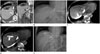
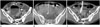
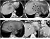
Of 21 abscesses, postoperative and non-postoperative abscesses were 9 (42.9%) and 12 (57.1%), respectively. Postoperative abscesses developed after the following operations: subtotal gastrectomy (n = 2), subtotal pancreatectomy (n = 1), hepatectomy (n = 3; 1 right hepatectomy and 2 left hepatectomy), laparoscopic cholecystectomy with left hepatectomy (n = 1), left hemicolectomy (n = 1), and distal pancreatectomy (n = 1). The location of the 21 abscesses were subphrenic (n = 7; 3 right, 4 left) (Fig. 1), pelvic (n = 5) (Fig. 2), periappendiceal (n = 3), peripancreatic (n = 2), liver (n = 2) (Fig. 3), pararenal (n = 1), and subhepatic (n = 1). Direct skin puncture for access was done in 18 cases. In 3 cases, the surgical drain or fistula opening was used as an access route. In case of direct puncture, the mean depth from skin puncture site to the abscess was 5.37 ± 1.97 cm.
The causes of inaccessibility were: narrow safe window due to adjacent or overlying organs (n = 9), irregularly dispersed abscess having a remote main area (n = 7), deep seated abscess with poor sonographic visualization (n = 4), and remote abscess from previously inserted surgical drain (n = 1). The details of inaccessibility are described in Table 1.
During the drainage procedure, a total of 29 CBCTs were performed. Multiple sessions of CBCT was performed in 6 abscesses. CBCT was performed thrice in 2 of the abscesses. For the other 4 abscesses, 2 sessions of CBCTs were performed in each. Cumulative air kerma and dose-area product of CBCT were 21.62 ± 5.41 mGy and 9179.87 ± 2337.70 mGycm2, respectively.
Technical and clinical success of the drainage was 20 (95.5%) and 21 (100%), respectively. In the 1 case with technical failure, catheter insertion into the main portion of the abscess failed due to the irregular dispersion of the abscess, despite a successful needle localization. However, complete drainage through communication of abscess locules culminated in a clinical success. Clinical success was achieved in all the 21 (100%) cases. Three patients underwent surgical treatment for an underlying disease, and the drainage catheter was removed during the surgery.
There were no complications during the drainage procedures.
DISCUSSION
US, fluoroscopy, computed tomography, or magnetic resonance imaging can be used for insertion of the drainage catheters during percutaneous procedures. Recent advancements of imaging modalities have facilitated more sophisticated needle or wire guidance. However, despite these improvements, some abscesses may appear to be inaccessible or undrainable due to an intrinsic limitation of imaging modality, unfavorable nature of the abscess, or topographic factors. In terms of imaging modality, US has been most widely used in percutaneous intervention for real-time guidance, but the acquired signal becomes weak as it travels deeper into the body. This results in the inevitable decrease of spatial resolution in imaging of deep seated lesions (19). In addition, for interventions in structures surrounded by air or air containing abscesses, US often cannot provide sufficient images because of artifacts (20). Unfavorable topographic factors are usually caused by anatomic distortions secondary to surgery, the surrounding organs, and difficult locations.
To overcome limitation due to access, procedures such as invasive transgressions of surrounding organs, or use of the surgical drain as an alternative access route rather than a straight access to the abscess, have previously been used (11). However, alternative approaches were not always possible because some major organs cannot be transgressed. In addition, surgical drains placed at the time of surgery are not always available.
The recently developed CBCT system enables imaging with wide z-axis coverage in 1 axial scan with ultra-high spatial resolution. Several studies for its potential application were previously reported in areas of clinical, preclinical, animal, and in vivo human imaging (12). Among the various CBCT systems, the C-arm based CBCT systems have a lower spatial resolution, as compared with CT gantry-based CBCT systems, since they are less mechanically stable and have limitations of C-arm rotation for data acquisition. However, C-arm based CBCT systems are particularity suitable for interventional and surgical application because they offer a better flexibility in orienting the detector around the patient, as compared to the closed CT gantry-based CBCT systems. In most cases, interventional procedures usually can be performed under C-arm fluoroscopy in the intervention room. However, cross sectional imaging, including CT scan, is sometimes needed to check the needle or guide wire position during the procedure. Therefore, application of CBCT system in C-arm fluoroscopy has been of great assistance in interventional procedures.
In our study, several factors contributed to inaccessibility to the abscess (Table 1). In such cases, US usually could not provide a sufficient view for access. However, we could achieve exact localization of the needle or guide wire by identifying and correcting their position on the basis of CBCT-derived three dimensional information. Especially in cases of postoperative abscess, CBCT was quite useful because the anatomic alterations had usually resulted in a poor sonic window, or there was presence of irregularly dispersed fluid collection.
In terms of the radiation dose of CBCT in our study, cumulative air kerma and dose-area product of CBCT were 21.62 ± 5.41 mGy and 9179.87 ± 2337.70 mGycm2, respectively. The radiation dose in our study cannot be directly compared to that of multidetector CT, because the CT dose index (CTDI) and the dose-length product (DLP) of multidetector CT cannot be used in CBCT (12). Although the total amount of radiation dose was more in CBCT, the post-procedural conventional CT to check the position of the drainage catheter, or re-intervention for reposition of drainage catheter, was avoided. Therefore, we believe that CBCT consequently reduces the possibility of additional radiation exposure by increasing the accuracy of the procedure.
In our study, the technical and clinical success of drainage was 20 (95.5%) and 21 (100%), respectively, with no complications related to the procedure. Without CBCT, some cases might be excluded or become surgical candidates. Therefore, although our cases were inaccessible abscesses, we had a high technical and clinical success rate. Even in patients with technical failure, CBCT provided the exact topographic information, and we could avoid catheter malposition outside the abscess cavity and finally achieve complete drainage of the abscess.
Our study had some limitations that warrant consideration. First, cases for CBCT were selected based on the decision of the attending radiologist. Therefore, our definition of poor accessibility was subjective and operator dependent. However, we believe that CBCT, though possibly unnecessary, can enhance the operator's confidence and accuracy of the procedure during drainage. Second, this study was limited by the small number of cases and retrospective nature. Therefore, further prospective studies with more cases are needed to confirm the clinical efficacy of CBCT in drainage of abscesses with poor accessibility.
In conclusion, CBCT is useful not only in targeting the inaccessible abscess, but the accuracy and success rates of drainage is also increased.
 XML Download