

 PDF
PDF ePub
ePub Citation
 Print
Print


INTRODUCTION
Hemangiomas account for less than 20% of all benign nasal cavity tumors (1). The majority of hemangiomas arise from the soft tissue of the nasal cavity, however, they can also arise from the bony structures. To the best of our knowledge, there are no prior case reports of intraosseous hemangiomas of the nasal septum. Here, we report one case of intraosseous cavernous hemangioma occurring in the nasal septum.
CASE REPORT
A 53-year-old woman without any significant medical history was admitted to our hospital for the surgical management of a mass in her nasal septum, which was found incidentally during brain magnetic resonance (MR) imaging at another hospital.
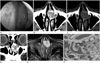
Nasal endoscopy revealed a large, reddish mass originating from the nasal septum (Fig. 1A). Computed tomography (CT) images revealed a well-defined, expansile, round mass within the anterior nasal septum. The mass showed multiple punctate calcifications, and remodeling of the adjacent bony structures was also seen (Fig. 1B, C). Contrast-enhanced CT images showed heterogeneous enhancement of the mass (Fig. 1D). Retrospective review of the original brain MR imaging revealed a well-defined mass within the anterior nasal septum. It showed high signal intensity with multiple internal low signal intensity foci representing calcifications on T2-weighted MR images (Fig. 1E). A contrast-enhanced MR imaging study was not performed. The mass was thought to be a chondroid tumor such as a chondroma, osteochondroma, or chondrosarcoma. It was completely excised via endoscopic surgery. Pathological examination revealed proliferation of small to intermediate-sized and dilated blood vessels interspersed among mature bone trabeculae (Fig. 1F). The final diagnosis was intraosseous cavernous hemangioma of the nasal septum.
DISCUSSION
Hemangiomas can occur as solitary bone lesions. Intraosseous hemangiomas in the head and neck are most commonly found in the skull, mandible, nasal bones, and cervical vertebrae. Although intraosseous hemangiomas of the sinonasal cavity are rare, several cases of nasal intraosseous hemangiomas arising from the inferior turbinate, middle turbinate, and paranasal areas have been reported (1234). The current case, to our knowledge, is the first report of intraosseous hemangioma involving the nasal septum.
Both CT and MR imaging are useful modalities for evaluating lesion characteristics in the sinonasal cavity. Prior literature has suggested that nasal intraosseous hemangiomas present as osteolytic masses with coarse internal trabeculation on CT, which is referred to as a soap bubble or honeycomb appearance (1234). MR imaging of these tumors demonstrates low signal intensity on T1-weighted images and high signal intensity on T2-weighted images with strong or globular enhancement (2). These findings are also similar to those of intraosseous hemangiomas elsewhere in the body (56).
As a wide variety of conditions can arise in the sinonasal cavity, and since their imaging findings are not usually pathognomonic, various tumorous lesions should be included in the differential diagnoses of a nasal cavity mass. Benign or malignant chondroid tumors arising within the nasal cavity tend to be lytic bone masses with densely scattered or ring and arc calcifications on CT (7), therefore, internal coarse trabeculation of hemangioma can mimic calcification of a chondroid tumor in the nasal cavity on imaging. Lobular capillary hemangioma, formerly known as pyogenic granuloma, is a benign soft tissue tumor characterized by rapid growth and a friable surface. It can occur in the anterior nasal septum in the area of the Kiesselbach plexus. Previous studies have reported that nasal trauma and pregnancy can be possible predisposing factors of lobular capillary hemangioma in the nasal septum (7). It usually appears as an intensely enhancing mass with an iso- to hypoattenuating cap and frequent adjacent bony changes (8). Benign or malignant minor salivary gland tumors can also arise at the nasal septum (79). For instance, a pleomorphic adenoma tends to be a multilobular tumor with curvilinear enhancement and small non-enhancing foci in the nasal septum. In addition, various other tumorous conditions should be included in the differential diagnoses of a nasal cavity mass, such as carcinoma, neuroendocrine tumor, schwannoma, angiofibroma, and Pindborg tumor (10).
En bloc resection of the tumor is the treatment of choice, and radiation therapy is another treatment option for intraosseous hemangioma (2).
Intraosseous hemangioma of the nasal cavity is a rare benign tumor. However, as it usually reveals relatively characteristic imaging findings such as an osteolytic mass with enhancement and internal coarse trabeculation, it should be considered in the differential diagnoses of a nasal septal tumor.
 XML Download